Copyright
©The Author(s) 2022.
World J Clin Cases. Mar 26, 2022; 10(9): 2916-2922
Published online Mar 26, 2022. doi: 10.12998/wjcc.v10.i9.2916
Published online Mar 26, 2022. doi: 10.12998/wjcc.v10.i9.2916
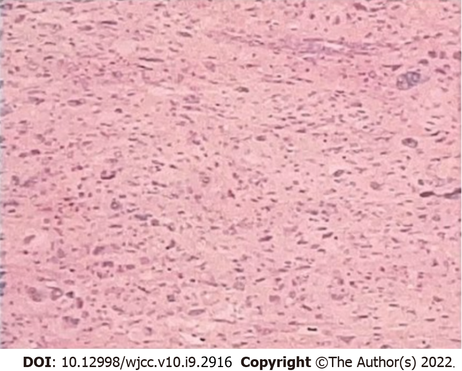
Figure 1 Postoperative pathological section (400 ×).
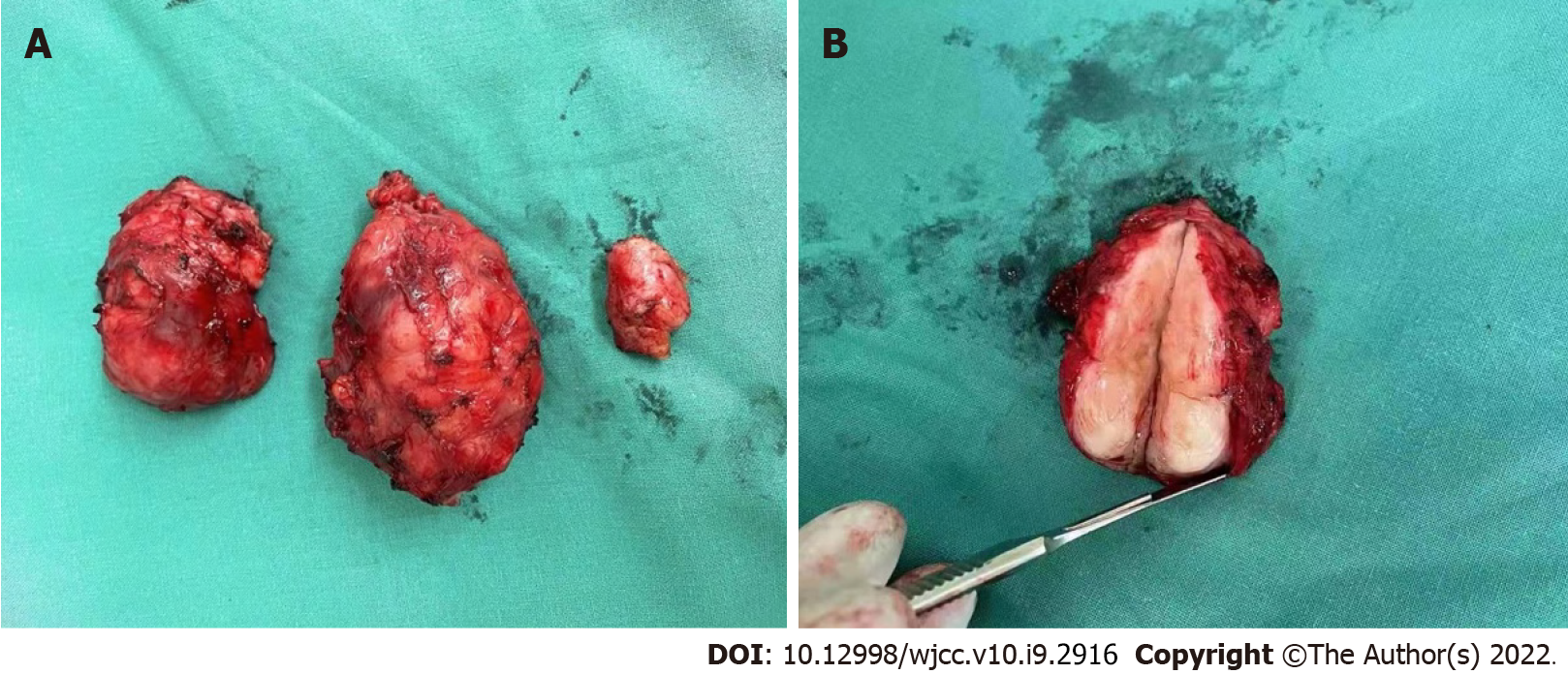
Figure 2 Three tumor specimens at the surgical removal site.
A: Tumor specimen; B: Maximum specimen profile.
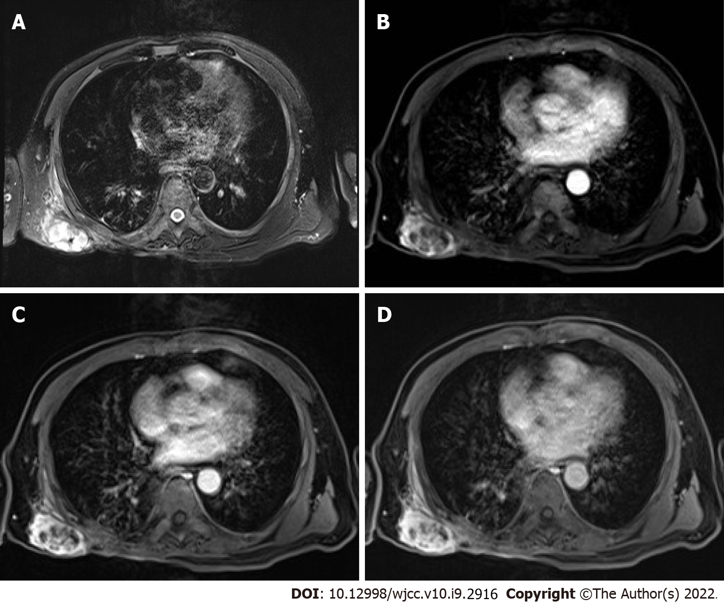
Figure 3 Preoperative chest MRI enhanced scan.
A: Cystic portion showed high signal in T2WI; B: Arterial phase, shows a multilocular solid and cystic lesion with slight enhancement on cyst wall; C: Venous phase; D: Delay period.
- Citation: Zhou YT, Wang RY, Zhang Y, Li DY, Yu J. Local hyperthermia combined with chemotherapy for the treatment of multiple recurrences of undifferentiated pleomorphic sarcoma: A case report. World J Clin Cases 2022; 10(9): 2916-2922
- URL: https://www.wjgnet.com/2307-8960/full/v10/i9/2916.htm
- DOI: https://dx.doi.org/10.12998/wjcc.v10.i9.2916


