Copyright
©The Author(s) 2022.
World J Clin Cases. Mar 26, 2022; 10(9): 2829-2835
Published online Mar 26, 2022. doi: 10.12998/wjcc.v10.i9.2829
Published online Mar 26, 2022. doi: 10.12998/wjcc.v10.i9.2829
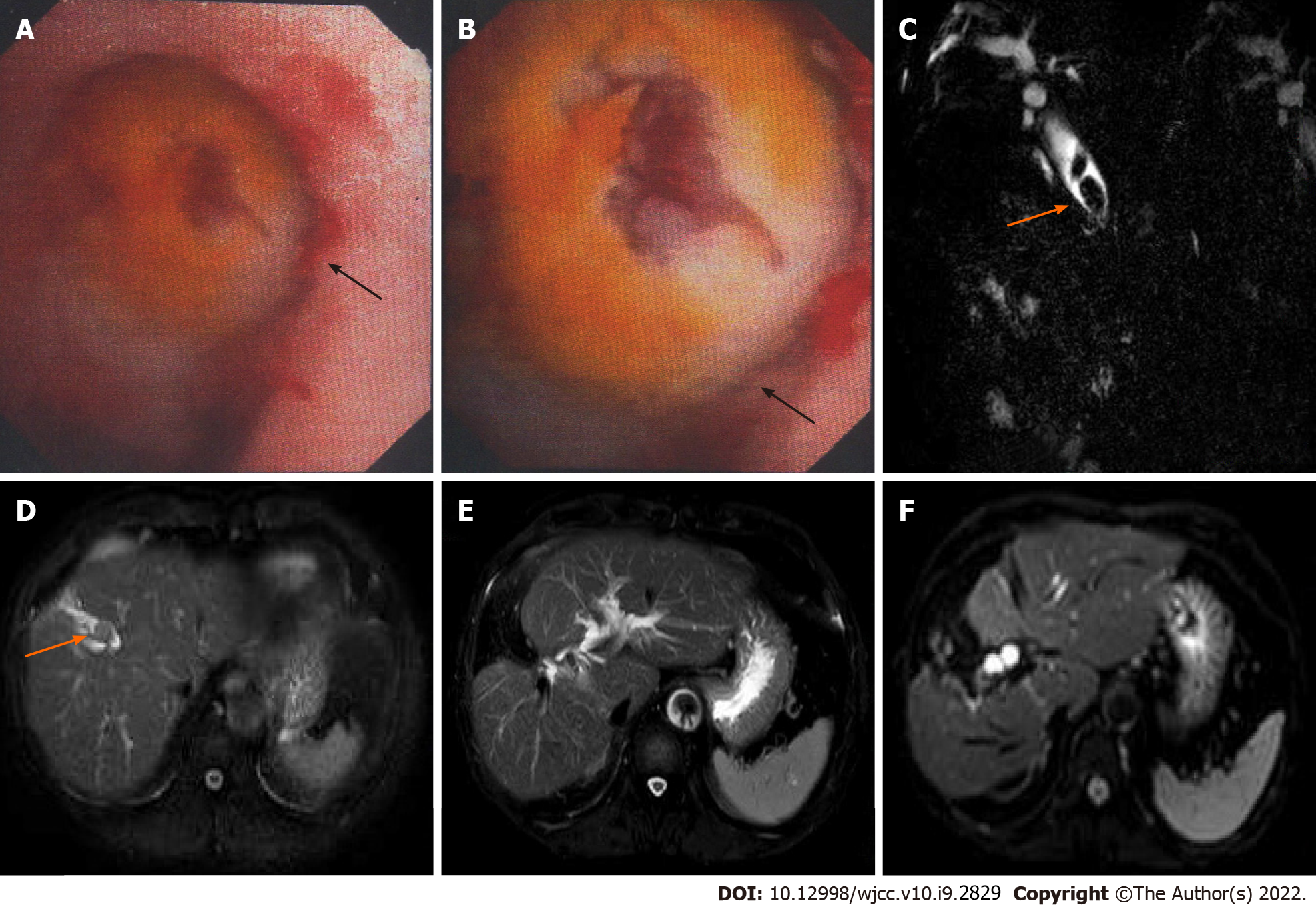
Figure 1 Magnetic resonance imaging findings and intraoperative choledochoscopy images of the patients.
A and B: Choledochoscopy showed a mass in the bile duct of the right anterior lobe during operation; C: Magnetic resonance cholangiopancreatography showed extrahepatic bile duct stones with bile duct dilatation before surgery; D: T2-weighted image showed intrahepatic bile duct stones with bile duct dilatation before surgery; E and F: T2-weighted image and diffusion-weighted imaging showed no tumor recurrence in the liver 72 mo after surgery.
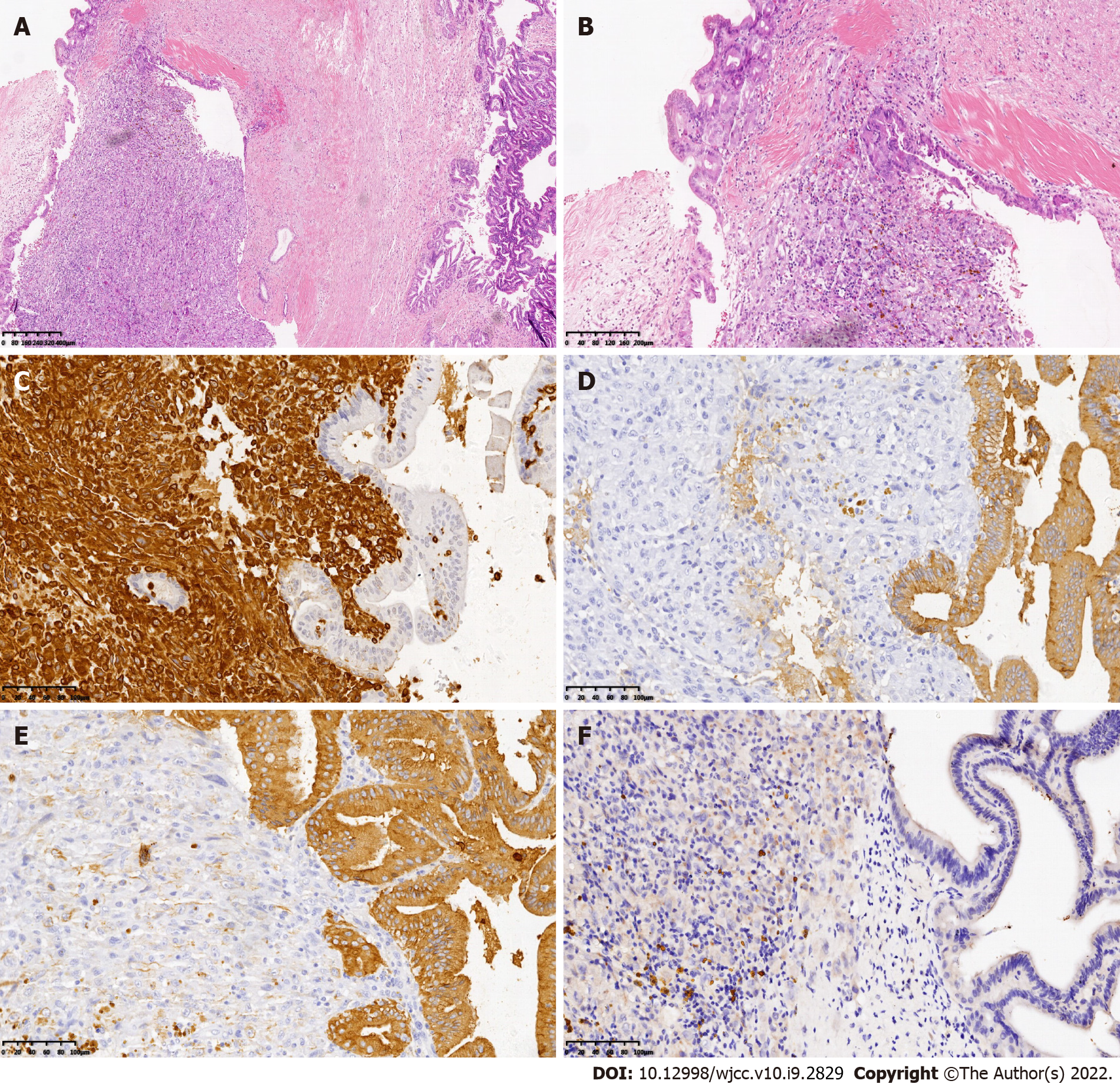
Figure 2 Histological characteristics and immunohistochemical images of the described patient with sarcomatoid intrahepatic cholangiocarcinoma.
A and B: The tumor consists of poorly differentiated intrahepatic cholangiocarcinoma (right) and spindle cells as the sarcomatoid component (left), which are intermingled (H&E stain, 40 × and 100 ×, respectively); C: Immunohistochemical staining showed sarcomatoid components stained with diffuse vimentin (vimentin staining, 200 ×); D: Immunohistochemistry showed positive expression of cytokeratin 19 (CK19) in cholangiocarcinoma cells (CK19 staining, 200 ×); E: Immunohistochemistry showed positive expression of pan-CK in cholangiocarcinoma cells (pan-CK staining, 200 ×); F: Immunohistochemistry showed negative expression of hepatocyte paraffin 1 (HepPar-1) in the cells (HepPar-1 staining, 200 ×). iCCA: Intrahepatic cholangiocarcinoma; H&E: Hematoxylin and eosin; CK19: Cytokeratin 19; pan-CK: Pan-cytokeratin; HepPar1: Hepatocyte paraffin 1.
- Citation: Feng JY, Li XP, Wu ZY, Ying LP, Xin C, Dai ZZ, Shen Y, Wu YF. Sarcomatoid intrahepatic cholangiocarcinoma with good patient prognosis after treatment with Huaier granules following hepatectomy: A case report. World J Clin Cases 2022; 10(9): 2829-2835
- URL: https://www.wjgnet.com/2307-8960/full/v10/i9/2829.htm
- DOI: https://dx.doi.org/10.12998/wjcc.v10.i9.2829


