Copyright
©The Author(s) 2022.
World J Clin Cases. Mar 6, 2022; 10(7): 2268-2274
Published online Mar 6, 2022. doi: 10.12998/wjcc.v10.i7.2268
Published online Mar 6, 2022. doi: 10.12998/wjcc.v10.i7.2268
Figure 1 Enhanced computed tomography of the abdomen of patient 4.
A: The ampullary mass shown by the orange arrow protrudes into the duodenal cavity, and the arterial phase shows uneven enhancement; B: The tumor caused significant dilation of the bile ducts and pancreatic ducts inside and outside the liver; C: Postoperative liver metastases indicated by orange arrows.
Figure 2 Enhanced magnetic resonance imaging of the abdomen and duodenoscopy of patient 3.
A: The orange arrow shows an oval mass in the ampulla, which shows high signal intensity on diffusion weighted imaging and the boundary is still clear; B: After the mass is enhanced, it shows uneven ring enhancement; C: An irregular ulcer in the ampulla was seen under the endoscopy, with a crater-like bulge around it.
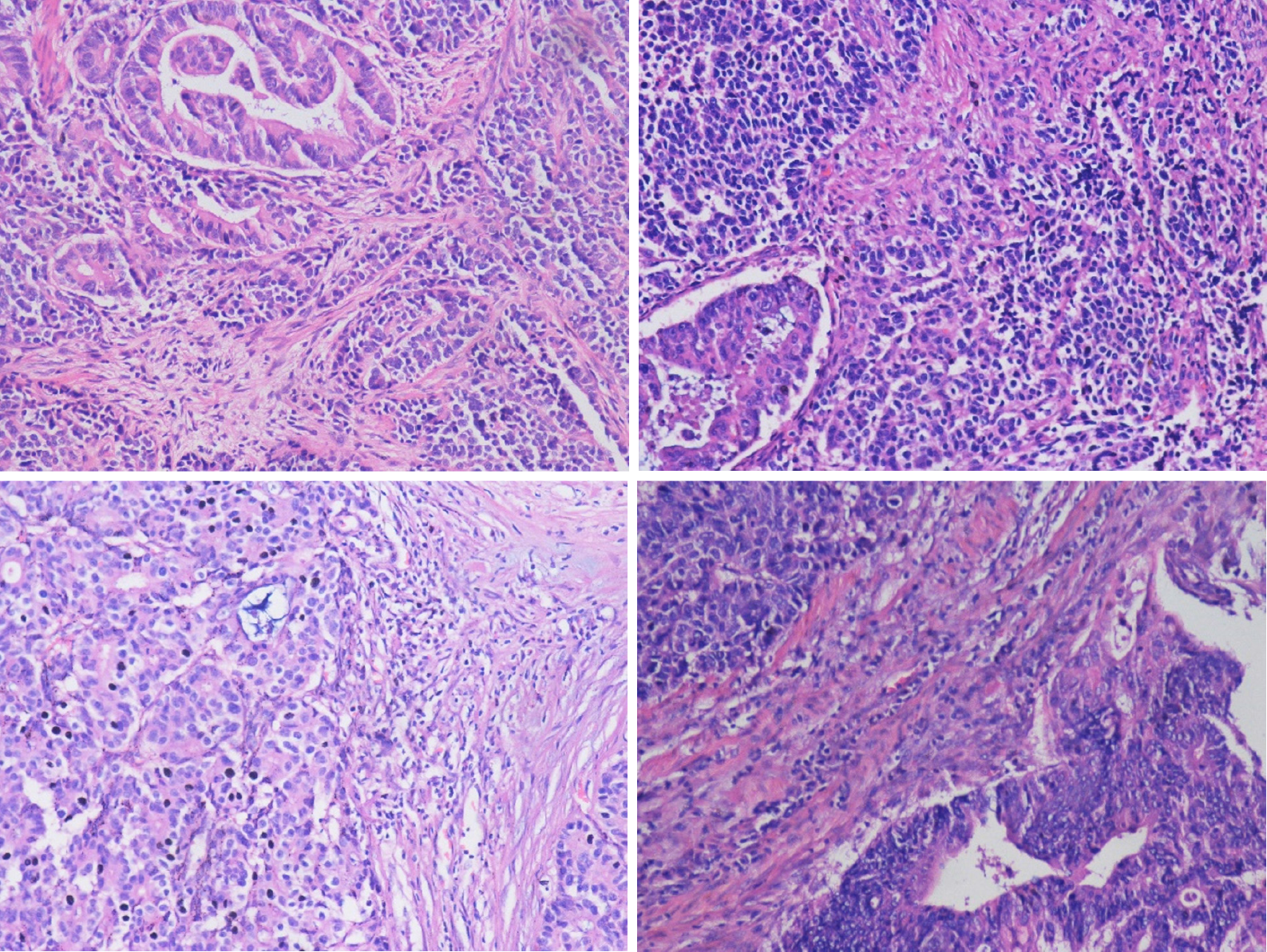
Figure 3 Light microscope images of hematoxylin-eosin stained lesion sections of 4 patients.
Original magnification: × 200.
Figure 4 Light microscope image with immunohistochemical staining.
A: CD56+; B: CgA+; C: Syn+. Original magnification: × 200.
- Citation: Wang Y, Zhang Z, Wang C, Xi SH, Wang XM. Mixed neuroendocrine-nonneuroendocrine neoplasm of the ampulla: Four case reports. World J Clin Cases 2022; 10(7): 2268-2274
- URL: https://www.wjgnet.com/2307-8960/full/v10/i7/2268.htm
- DOI: https://dx.doi.org/10.12998/wjcc.v10.i7.2268


