Copyright
©The Author(s) 2022.
World J Clin Cases. Feb 16, 2022; 10(5): 1630-1638
Published online Feb 16, 2022. doi: 10.12998/wjcc.v10.i5.1630
Published online Feb 16, 2022. doi: 10.12998/wjcc.v10.i5.1630
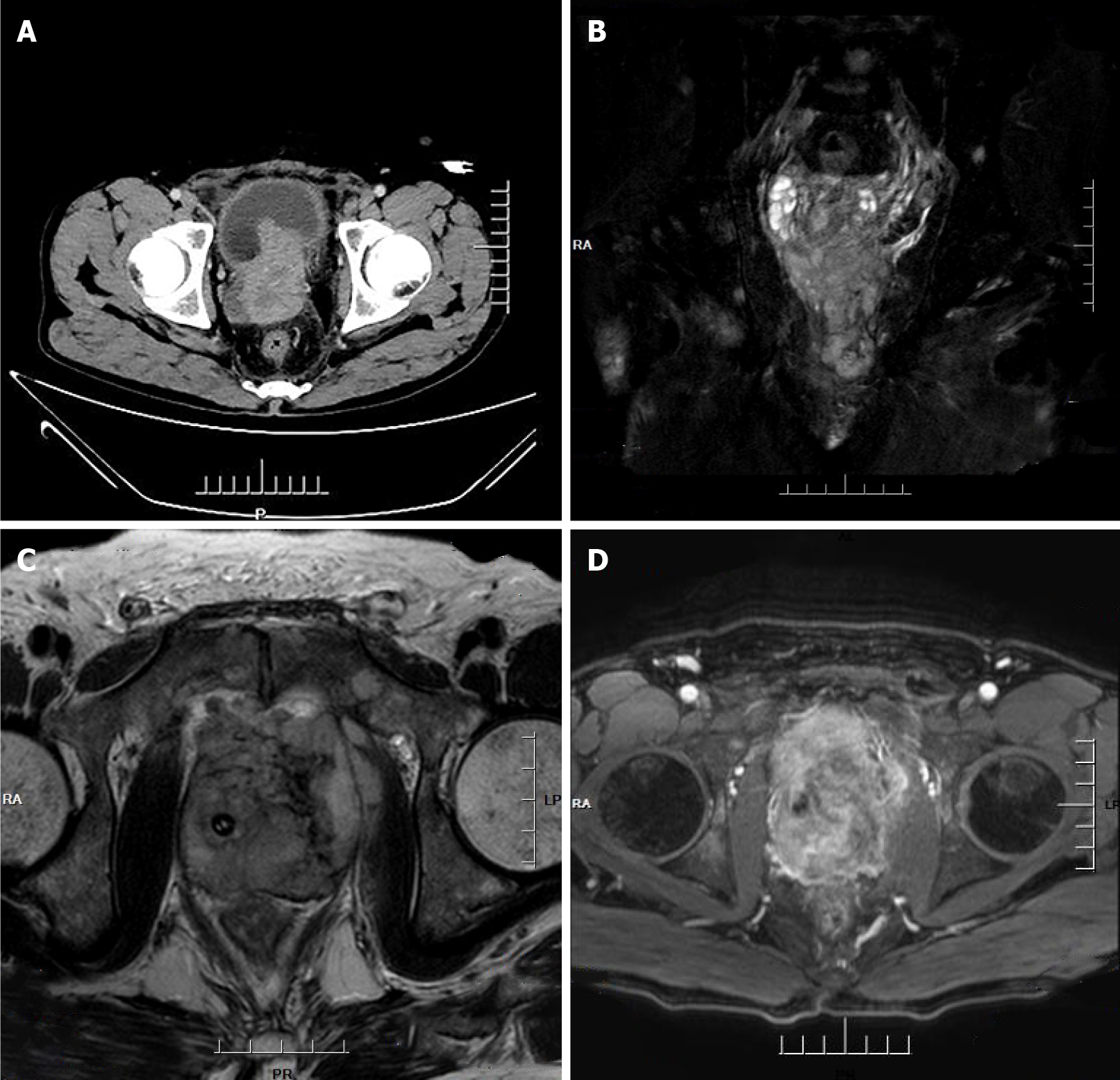
Figure 1 Computed tomography imaging and magnetic resonance imagining.
A: Computed tomography imaging of the abdomen: Heterogeneous density within the prostate and heterogeneous enhancement after enhancement, suggesting possible prostate cancer, possible pelvic lymph node metastasis, pelvic floor fascia, and rectal wall and seminal vesicle invasion; B-D: Prostate magnetic resonance imagining: Mixed signals in the prostate, with possible prostate tumor invasion of the lower posterior bladder wall, rectal mesentery, and bilateral seminal vesicles, with multiple lymph node metastases in the pelvis.
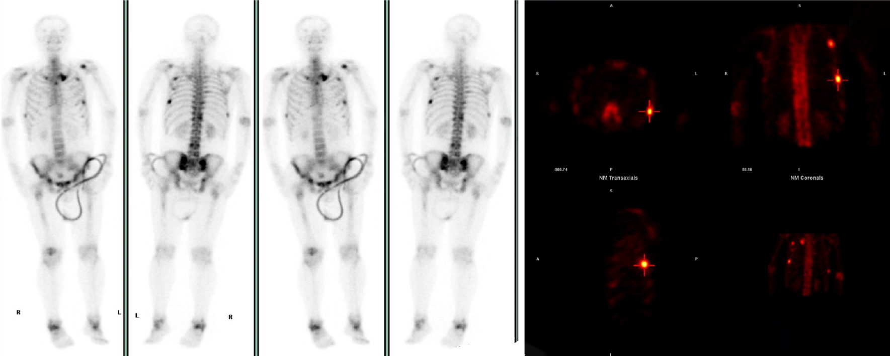
Figure 2
Whole-body bone scan: Abnormally active bone metabolism in the left sternoclavicular joint, multiple ribs, T12, L4, L5 vertebrae, and right acetabulum.
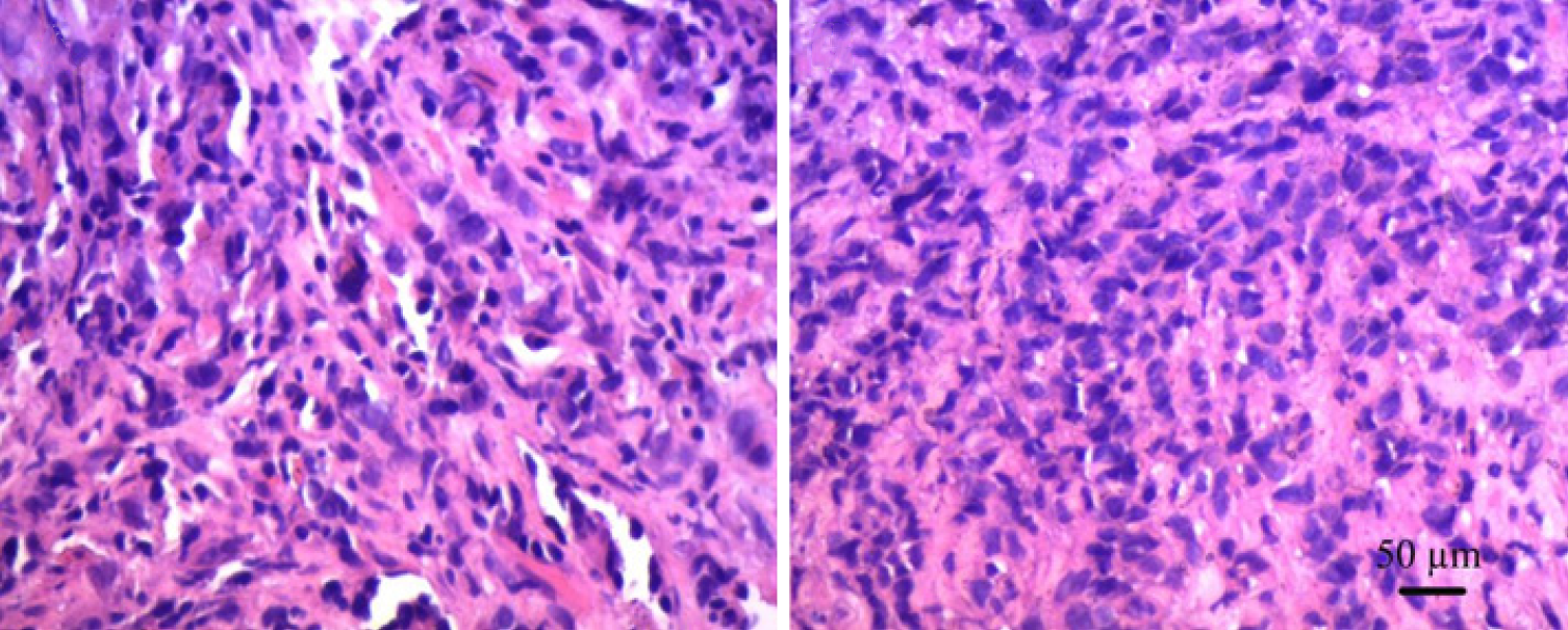
Figure 3
HE × 200 ×; tumor cells arranged in strips and sheets; tumor cells appear oval or spindle-shaped.
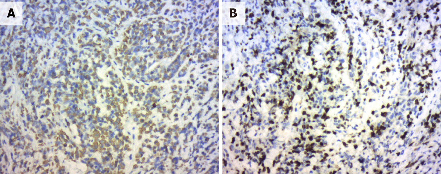
Figure 4 Immunohistochemical.
A: SP method immunohistochemical staining for CK broadly focally positive; B: SP method immunohistochemical staining for Ki67 (50%+).
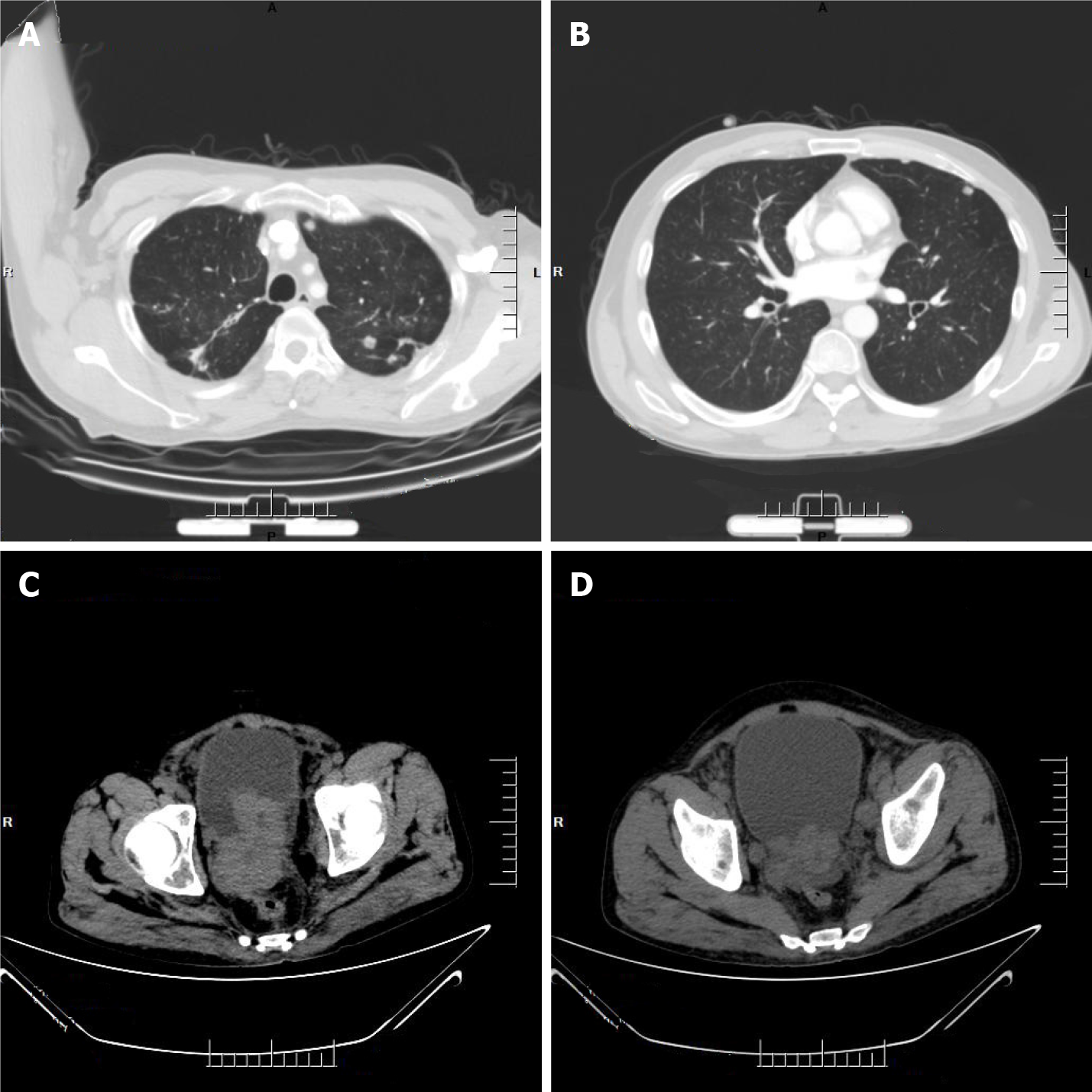
Figure 5 Computed tomography.
A and B: Computed tomography (CT) imaging of the lung: Multiple nodular shadows in both lungs, with possible Small-cell carcinoma of the prostate metastases; C and D: CT imaging of the abdomen: An irregular enlargement of Small-cell carcinoma of the prostate showing a tendency to infiltrate, bilateral involvement of the inner segment of the ureteral bladder wall, dilatation and fluid retention in the urinary tract, and multiple bone destruction in the pelvis.
- Citation: Shi HJ, Fan ZN, Zhang JS, Xiong BB, Wang HF, Wang JS. Small-cell carcinoma of the prostate with negative CD56, NSE, Syn, and CgA indicators: A case report. World J Clin Cases 2022; 10(5): 1630-1638
- URL: https://www.wjgnet.com/2307-8960/full/v10/i5/1630.htm
- DOI: https://dx.doi.org/10.12998/wjcc.v10.i5.1630


