
















©The Author(s) 2022.
World J Clin Cases. Feb 16, 2022; 10(5): 1630-1638
Published online Feb 16, 2022. doi: 10.12998/wjcc.v10.i5.1630
Published online Feb 16, 2022. doi: 10.12998/wjcc.v10.i5.1630
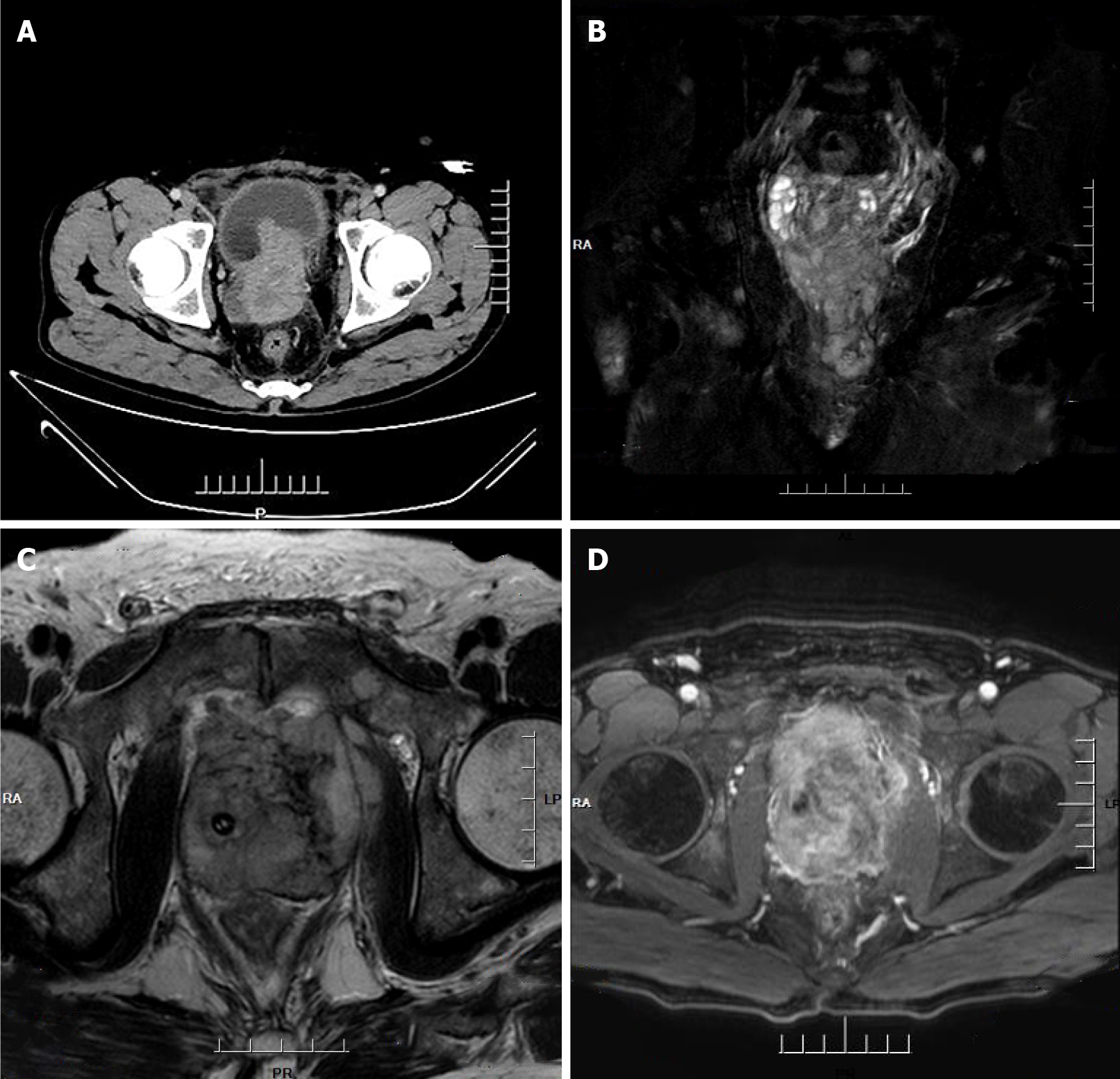
Figure 1 Computed tomography imaging and magnetic resonance imagining.
A: Computed tomography imaging of the abdomen: Heterogeneous density within the prostate and heterogeneous enhancement after enhancement, suggesting possible prostate cancer, possible pelvic lymph node metastasis, pelvic floor fascia, and rectal wall and seminal vesicle invasion; B-D: Prostate magnetic resonance imagining: Mixed signals in the prostate, with possible prostate tumor invasion of the lower posterior bladder wall, rectal mesentery, and bilateral seminal vesicles, with multiple lymph node metastases in the pelvis.
- Citation: Shi HJ, Fan ZN, Zhang JS, Xiong BB, Wang HF, Wang JS. Small-cell carcinoma of the prostate with negative CD56, NSE, Syn, and CgA indicators: A case report. World J Clin Cases 2022; 10(5): 1630-1638
- URL: https://www.wjgnet.com/2307-8960/full/v10/i5/1630.htm
- DOI: https://dx.doi.org/10.12998/wjcc.v10.i5.1630