Copyright
©The Author(s) 2022.
World J Clin Cases. Dec 16, 2022; 10(35): 13064-13073
Published online Dec 16, 2022. doi: 10.12998/wjcc.v10.i35.13064
Published online Dec 16, 2022. doi: 10.12998/wjcc.v10.i35.13064
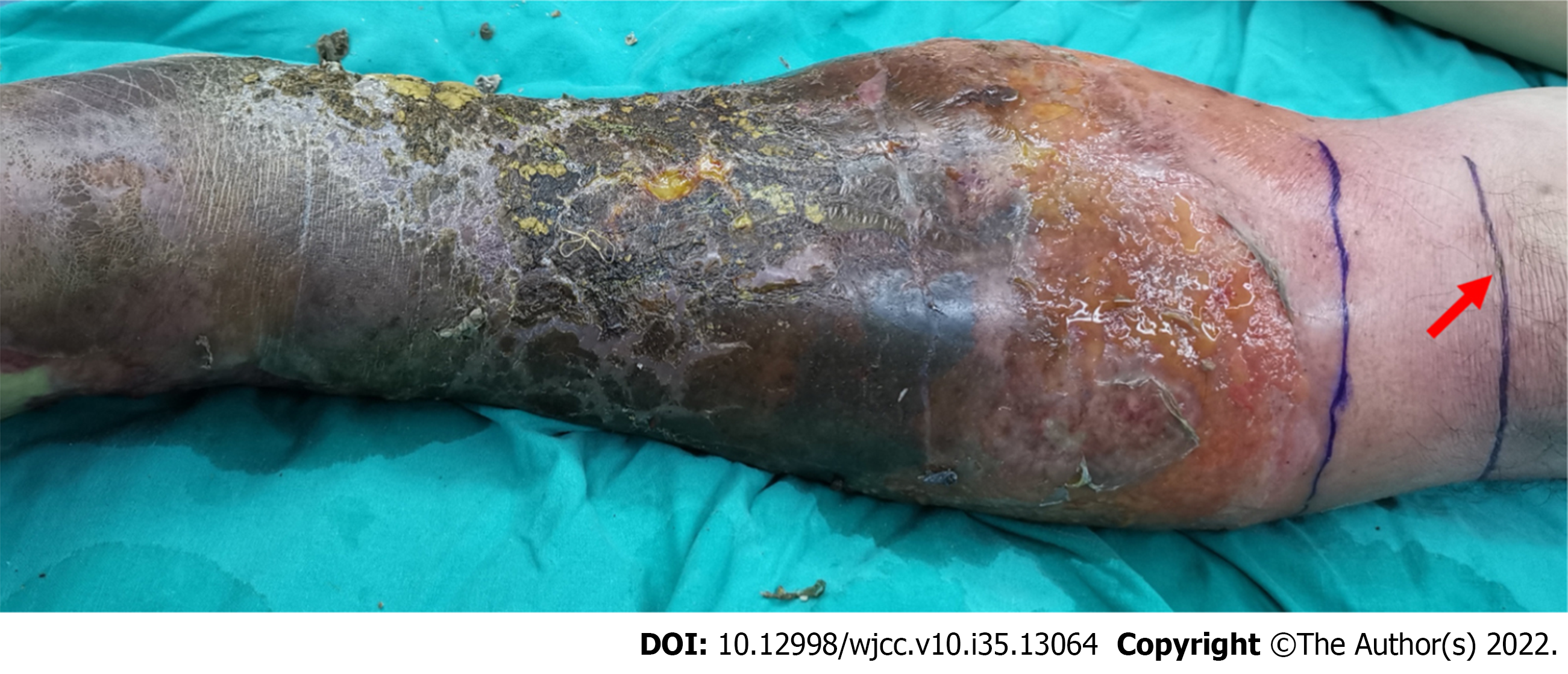
Figure 1 Skin lesions of the left lower limb on admission.
Figure 2 Left lower extremity skin lesions at different stages of treatment and postoperative pathological examination results.
A: Image of left lower limb after enlarged debridement; B: The pathological findings of surgical specimens suggested the formation of soft tissue abscess; C: Image of left lower limb flap after transplantation; D: Left lower extremity flap transplantation.
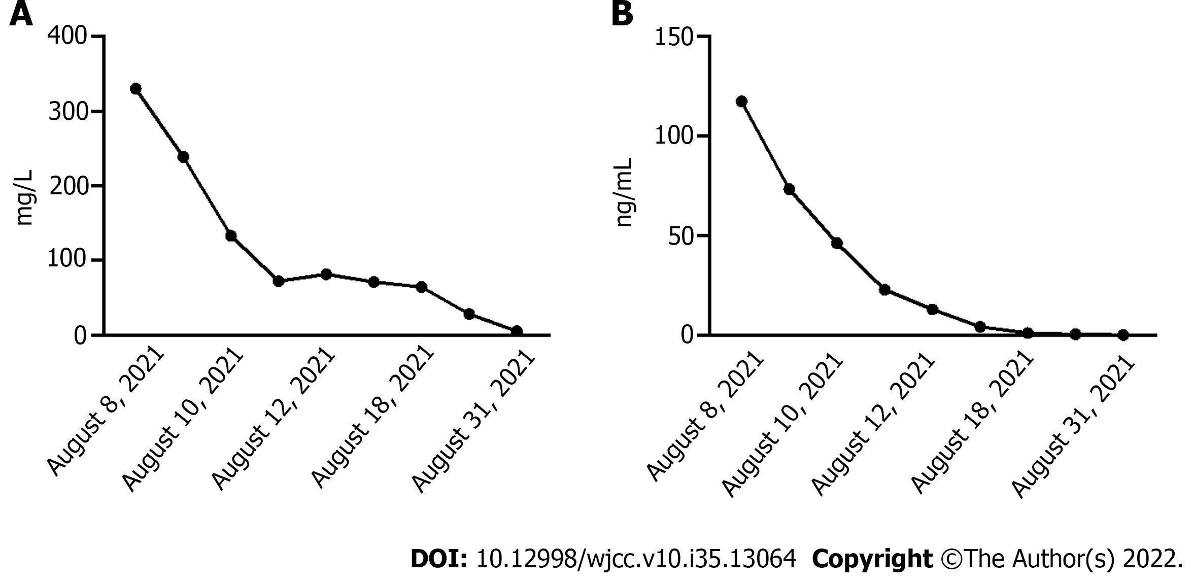
Figure 3 Patient’s infection index.
A: C-reactive protein; B: Procalcitonin.
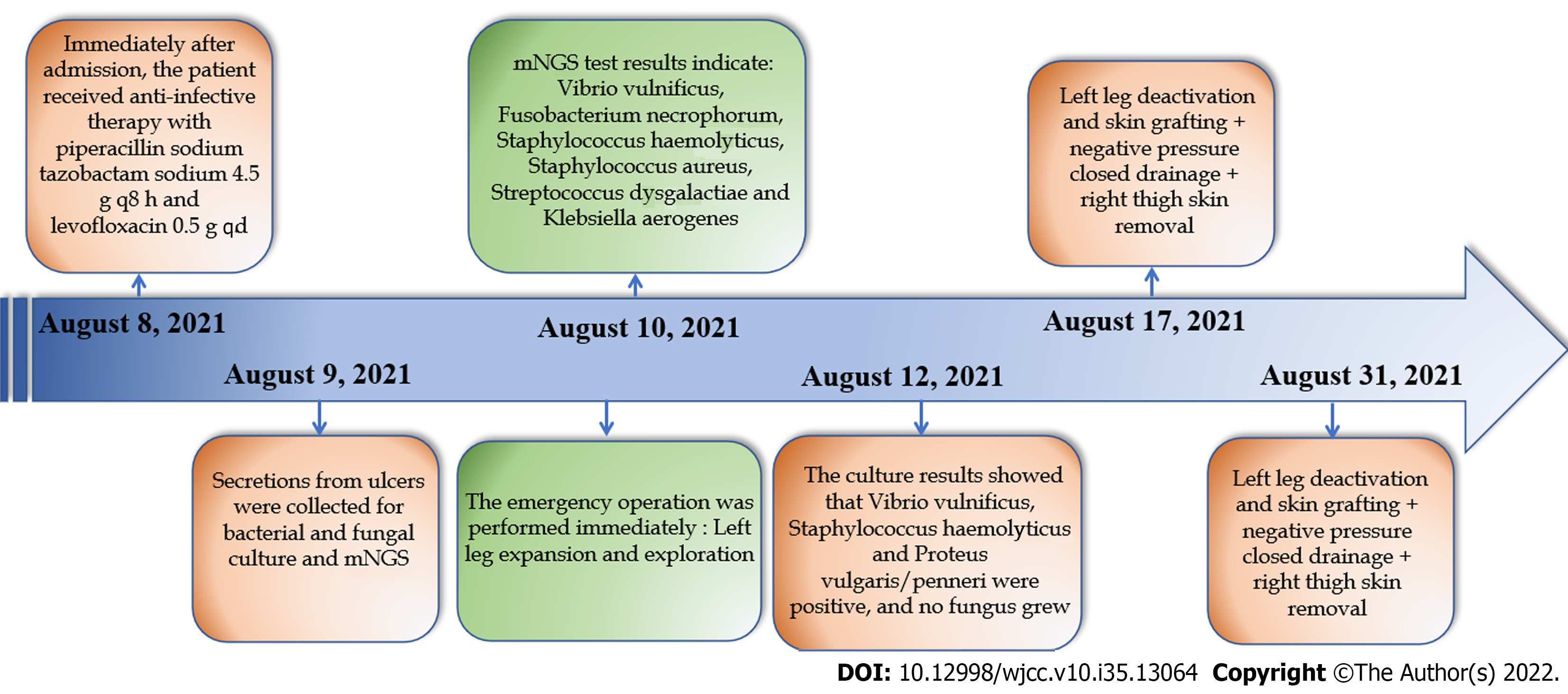
Figure 4 Flow charts of diagnosis and treatment.
mNGS: Metagenomics next-generation sequencing.
- Citation: Lu HY, Gao YB, Qiu XW, Wang Q, Liu CM, Huang XW, Chen HY, Zeng K, Li CX. Successful surgical treatment of polybacterial gas gangrene confirmed by metagenomic next-generation sequencing detection: A case report. World J Clin Cases 2022; 10(35): 13064-13073
- URL: https://www.wjgnet.com/2307-8960/full/v10/i35/13064.htm
- DOI: https://dx.doi.org/10.12998/wjcc.v10.i35.13064


