Copyright
©The Author(s) 2022.
World J Clin Cases. Dec 16, 2022; 10(35): 13044-13051
Published online Dec 16, 2022. doi: 10.12998/wjcc.v10.i35.13044
Published online Dec 16, 2022. doi: 10.12998/wjcc.v10.i35.13044
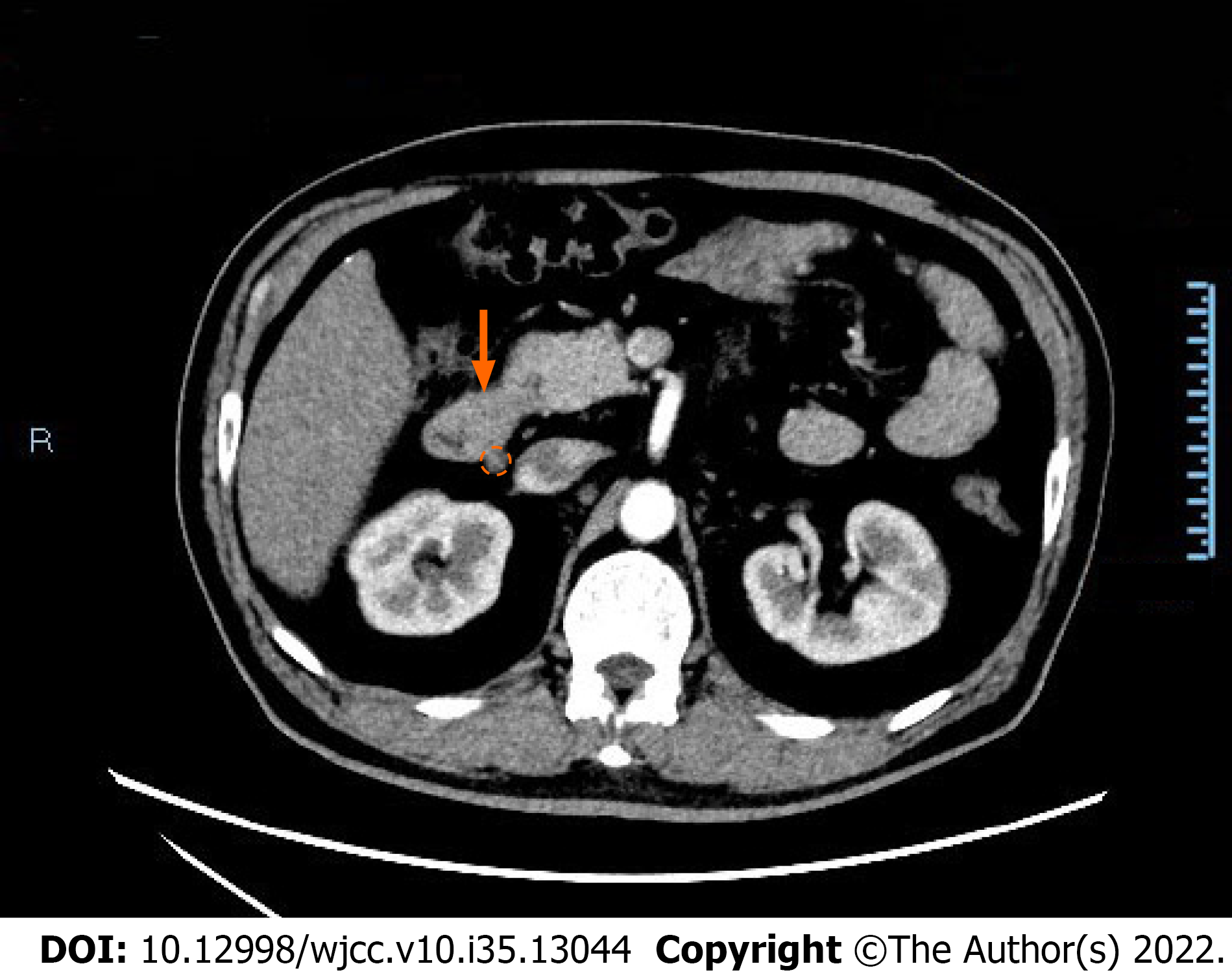
Figure 1 Contrast-enhanced computed tomography image showing abnormality of the descending duodenum and lymph nodes.
Focal thickening of the wall of the descending duodenum (orange arrow) and enlarged lymph nodes (orange dotted circle) are shown in the computed tomography image.
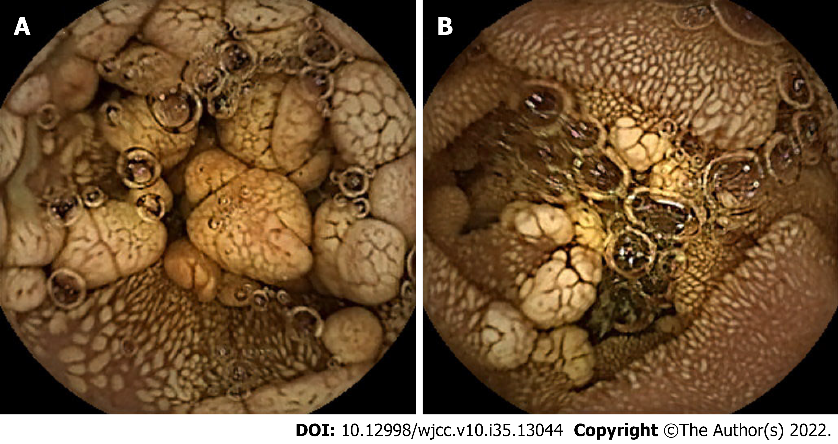
Figure 2 Capsule endoscopy images of multiple polypoid bulges in the duodenum and proximal jejunum.
A: Grayish-yellow polypoid bulges of various sizes were observed in the duodenum; B: Grayish-yellow polypoid bulges of various sizes were observed in the proximal jejunum.
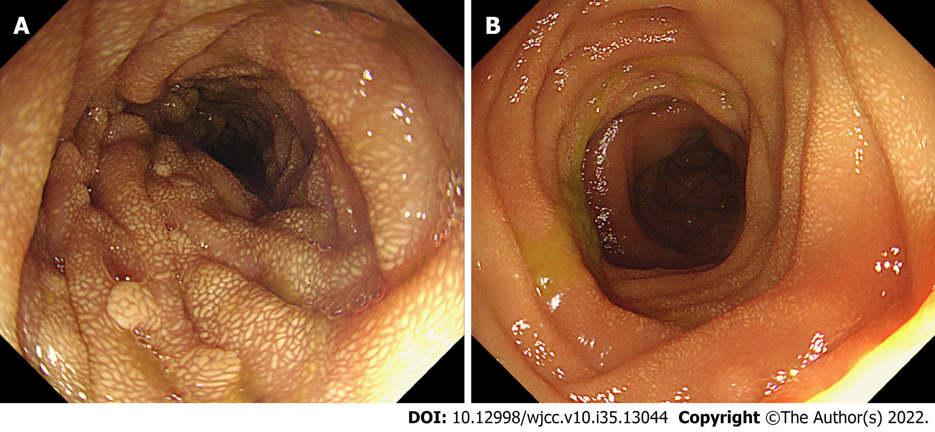
Figure 3 Endoscopic images of the jejunum during the operation.
A: Proximal jejunum; B: Distal jejunum. Multiple polyps were observed in the proximal jejunum and gradually disappeared in the distal jejunum. Whitish-yellow plaque-like changes were diffusely distributed both in the proximal and distal jejunum.
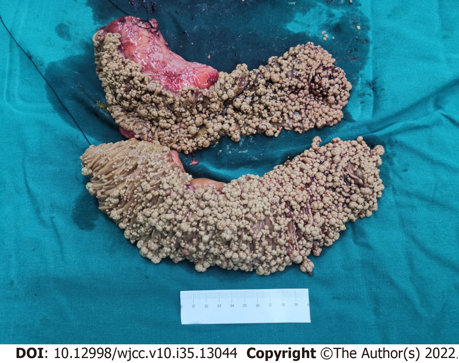
Figure 4 Specimens of the removed pancreatic head and small intestine.
The upper specimen included the pancreatic head (top left) and partial duodenum, and the lower specimen was the proximal jejunum. The mucosal surface of the intestine was covered with grayish-yellow polypoid bulges with various sizes ranging from the needle tip to 2 cm × 1 cm × 1 cm.
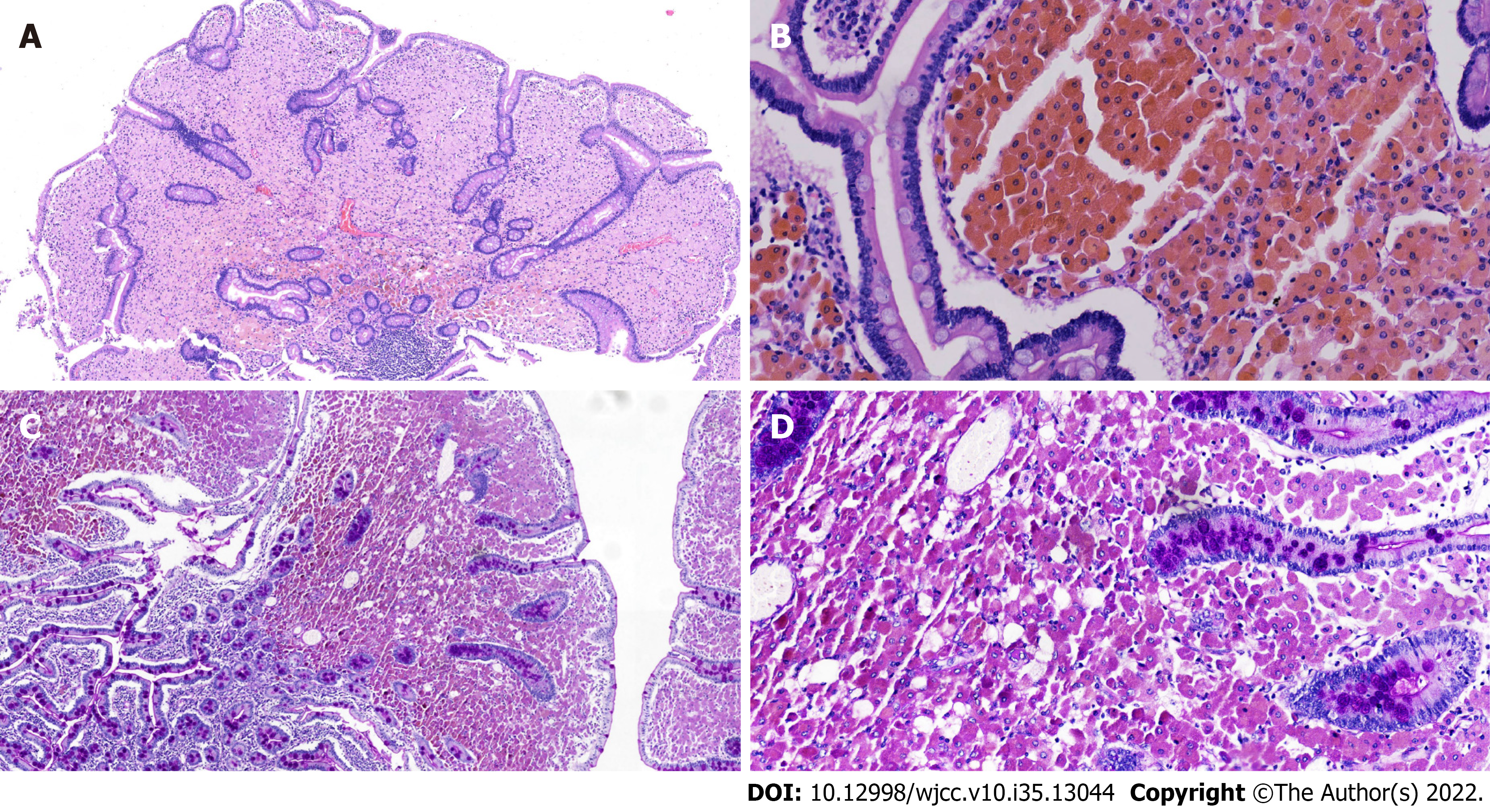
Figure 5 Histological section of a duodenum specimen.
Hematoxylin-eosin (HE) and periodic acid-Schiff (PAS) staining showed that many macrophages were aggregated in the lamina propria of the duodenum. A: HE staining at low magnification; B: HE staining at high magnification; C: Positive PAS staining at low magnification; D: Positive PAS staining at high magnification.
- Citation: Chen S, Zhou YC, Si S, Liu HY, Zhang QR, Yin TF, Xie CX, Yao SK, Du SY. Atypical Whipple’s disease with special endoscopic manifestations: A case report. World J Clin Cases 2022; 10(35): 13044-13051
- URL: https://www.wjgnet.com/2307-8960/full/v10/i35/13044.htm
- DOI: https://dx.doi.org/10.12998/wjcc.v10.i35.13044


