Copyright
©The Author(s) 2022.
World J Clin Cases. Dec 16, 2022; 10(35): 13044-13051
Published online Dec 16, 2022. doi: 10.12998/wjcc.v10.i35.13044
Published online Dec 16, 2022. doi: 10.12998/wjcc.v10.i35.13044
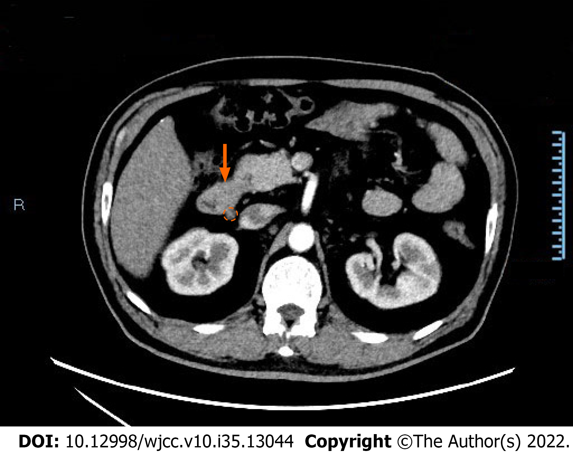
Figure 1 Contrast-enhanced computed tomography image showing abnormality of the descending duodenum and lymph nodes.
Focal thickening of the wall of the descending duodenum (orange arrow) and enlarged lymph nodes (orange dotted circle) are shown in the computed tomography image.
- Citation: Chen S, Zhou YC, Si S, Liu HY, Zhang QR, Yin TF, Xie CX, Yao SK, Du SY. Atypical Whipple’s disease with special endoscopic manifestations: A case report. World J Clin Cases 2022; 10(35): 13044-13051
- URL: https://www.wjgnet.com/2307-8960/full/v10/i35/13044.htm
- DOI: https://dx.doi.org/10.12998/wjcc.v10.i35.13044


