Copyright
©The Author(s) 2022.
World J Clin Cases. Nov 6, 2022; 10(31): 11585-11589
Published online Nov 6, 2022. doi: 10.12998/wjcc.v10.i31.11585
Published online Nov 6, 2022. doi: 10.12998/wjcc.v10.i31.11585
Figure 1 The centre of the plaque was slightly atrophic and depressed.
A: Left plantar skin lesions in patient with perspiration keratosis; B: There were scattered ring-shaped light-brown keratinizing papules at the bottom of the left foot, about 0.3-0.5 cm in diameter, partially fused, diffuse and banded, distributed along the B line; C: The surface was rough, the boundary was clear, and the skin in the center of the lesions was slightly sunken and atrophic.
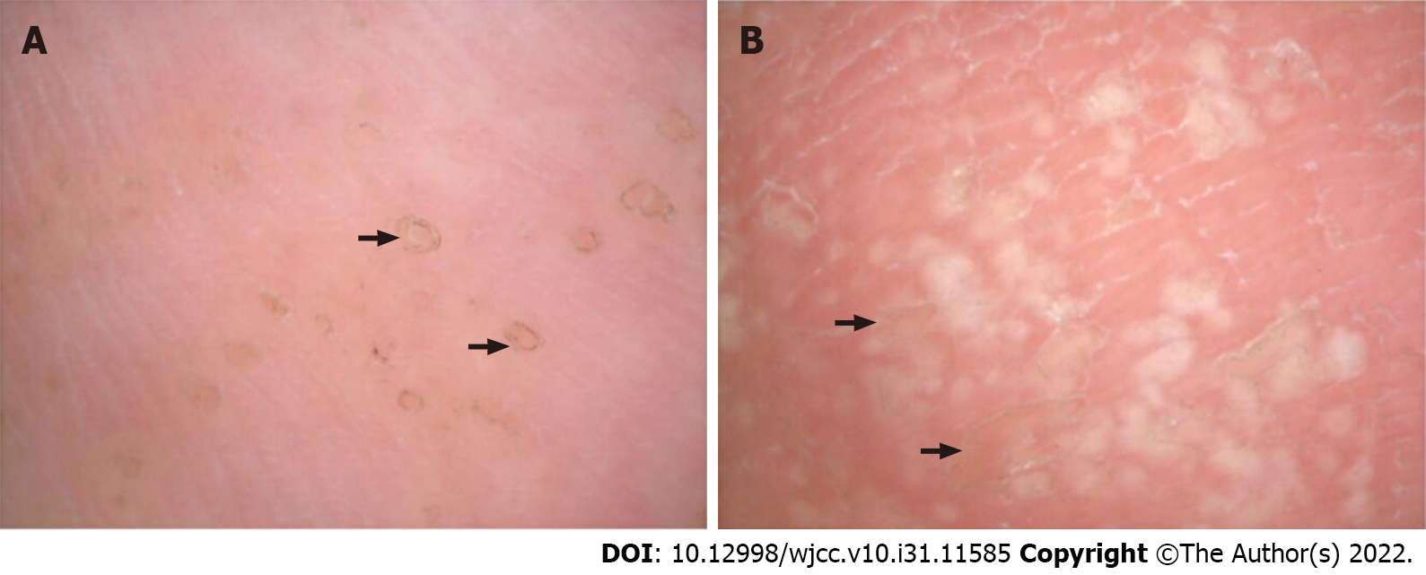
Figure 2 Dermoscopic manifestations of plantar skin lesions in porokeratosis.
A: The foot base is scattered in black brown ring structure, the edge of the dike; B: Irregular pale white hypopigmented areas were seen in the center of the skin lesions.
Figure 3 Pathological images of skin lesions of porokeratosis.
A: HE staining, ×100, B: HE staining, ×200, C: HE staining, ×400. Epidermis was hyperkeratosis with a high degree of hyperkeratosis, with columns of parakeratosis, thinning of the granular layer, thickening of the spinous layer, and a small amount of lymphocyte infiltration in the superficial dermis.
- Citation: Yang J, Du YQ, Fang XY, Li B, Xi ZQ, Feng WL. Linear porokeratosis of the foot with dermoscopic manifestations: A case report. World J Clin Cases 2022; 10(31): 11585-11589
- URL: https://www.wjgnet.com/2307-8960/full/v10/i31/11585.htm
- DOI: https://dx.doi.org/10.12998/wjcc.v10.i31.11585


