Copyright
©The Author(s) 2022.
World J Clin Cases. Nov 6, 2022; 10(31): 11523-11528
Published online Nov 6, 2022. doi: 10.12998/wjcc.v10.i31.11523
Published online Nov 6, 2022. doi: 10.12998/wjcc.v10.i31.11523
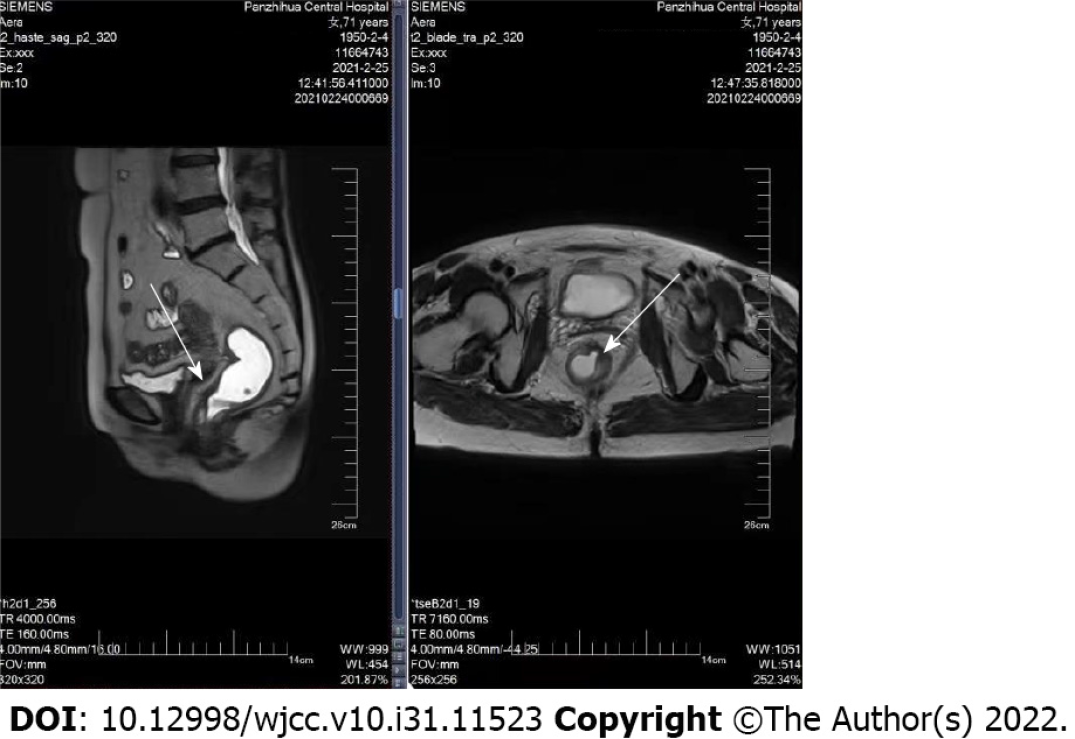
Figure 1 Rectal magnetic resonance imaging shows the area of lesion (white arrow).
There was no specific abdominal ascites and no mesenteric lymph node enlargement.
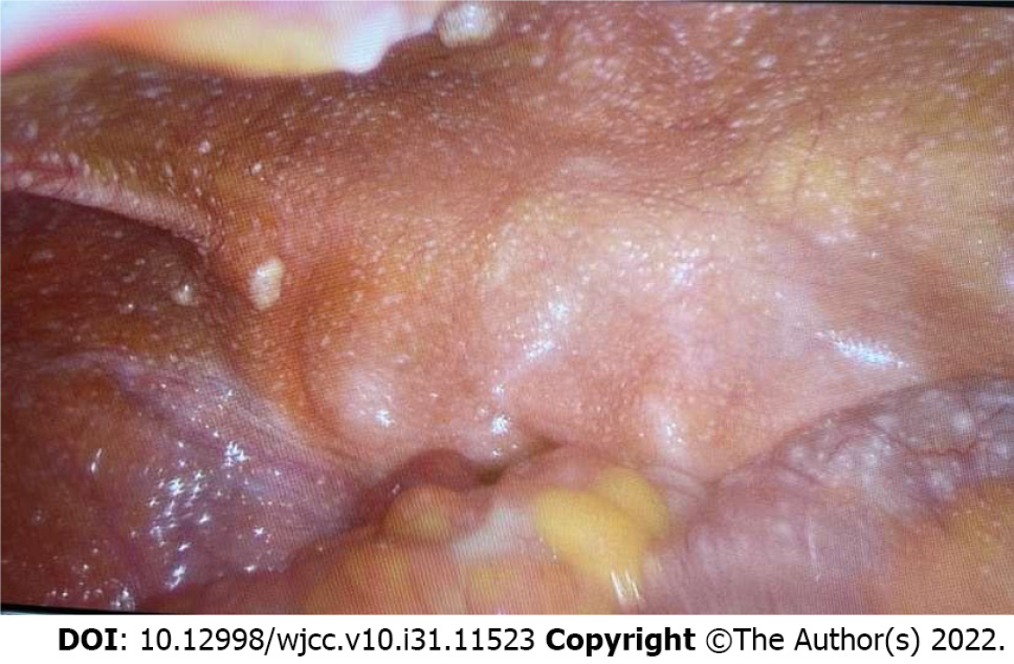
Figure 2 Disseminated peritoneal nodules detected during laparoscopy.
Representative biopsies were taken.
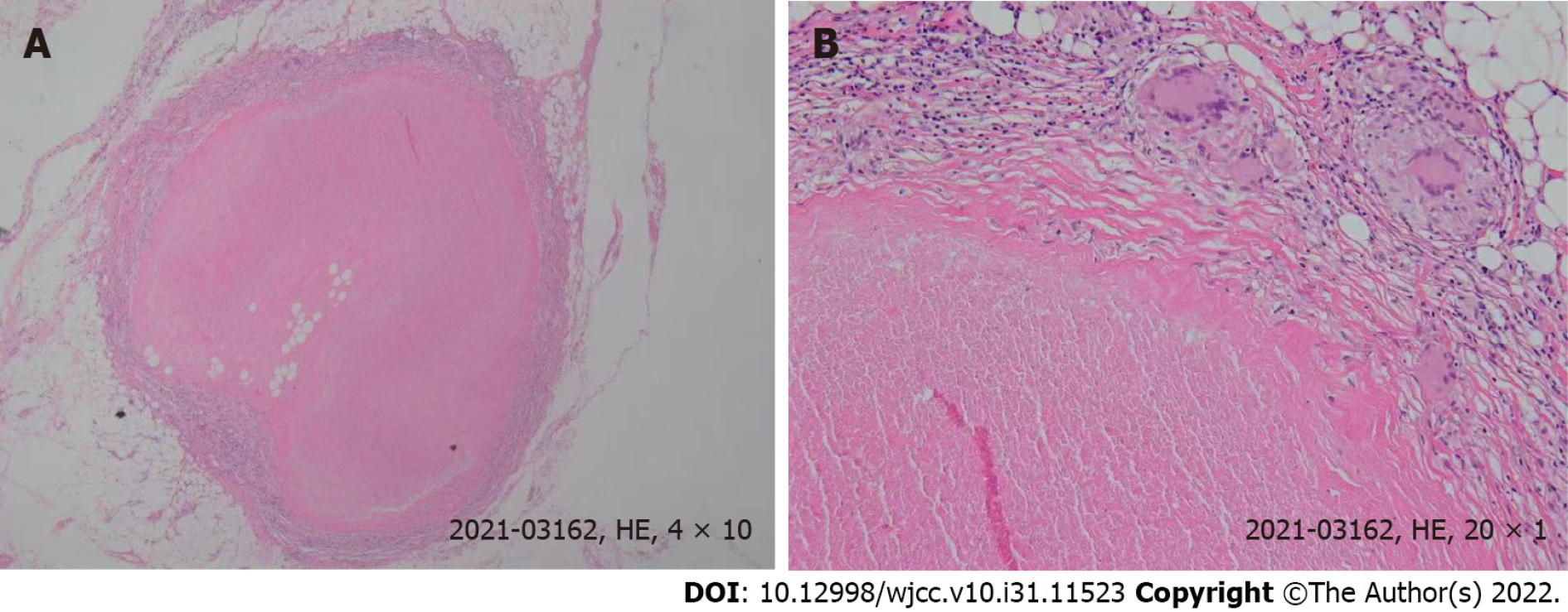
Figure 3 Pathologic findings.
A: Histopathological image of a biopsy, taken from the greater omentum, showing granulomas (hematoxylin and eosin stain, magnification 40 ×); B: Histopathological image of granulomas (hematoxylin and eosin stain, magnification 200 ×). HE: Hematoxylin and eosin.
- Citation: Liu PG, Chen XF, Feng PF. Rectal cancer combined with abdominal tuberculosis: A case report. World J Clin Cases 2022; 10(31): 11523-11528
- URL: https://www.wjgnet.com/2307-8960/full/v10/i31/11523.htm
- DOI: https://dx.doi.org/10.12998/wjcc.v10.i31.11523


