Copyright
©The Author(s) 2022.
World J Clin Cases. Oct 26, 2022; 10(30): 11172-11177
Published online Oct 26, 2022. doi: 10.12998/wjcc.v10.i30.11172
Published online Oct 26, 2022. doi: 10.12998/wjcc.v10.i30.11172
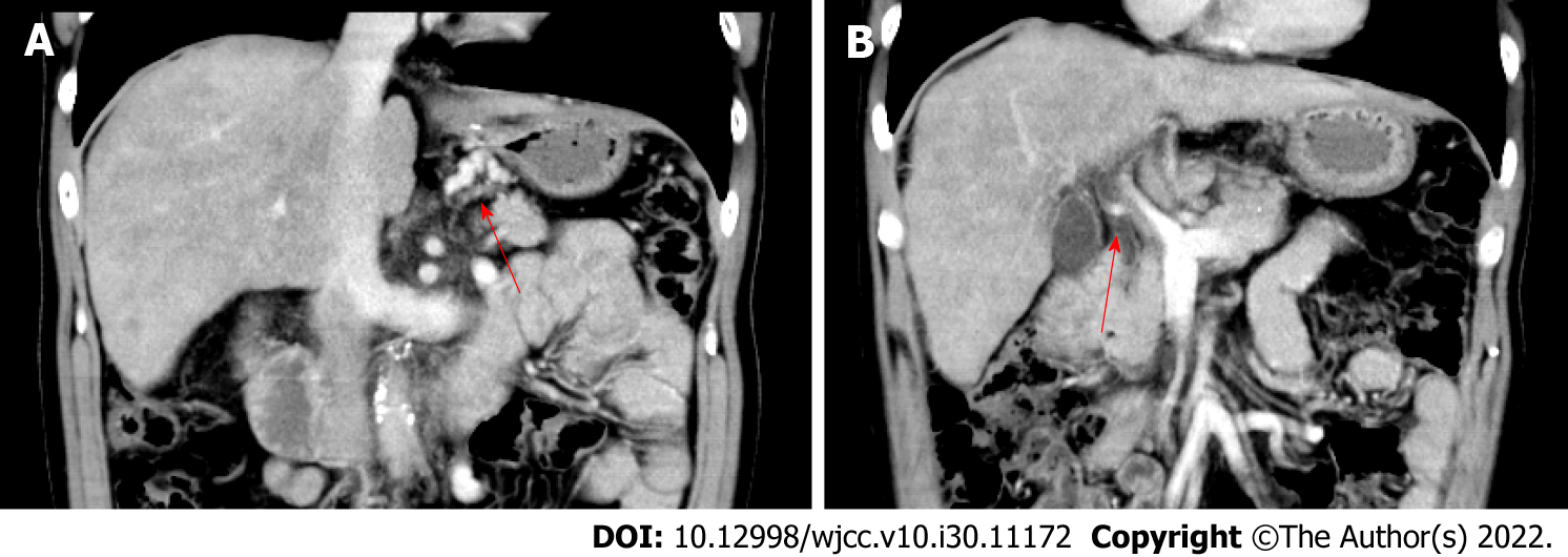
Figure 1 Contrast abdominal computed tomography revealed cirrhosis, esophageal and gastric fundus varicose veins, fundus-left renal shunt, and portal vein thrombosis.
A: Arrow shows portal vein thrombosis; B: Arrow shows esophagogastric varices.
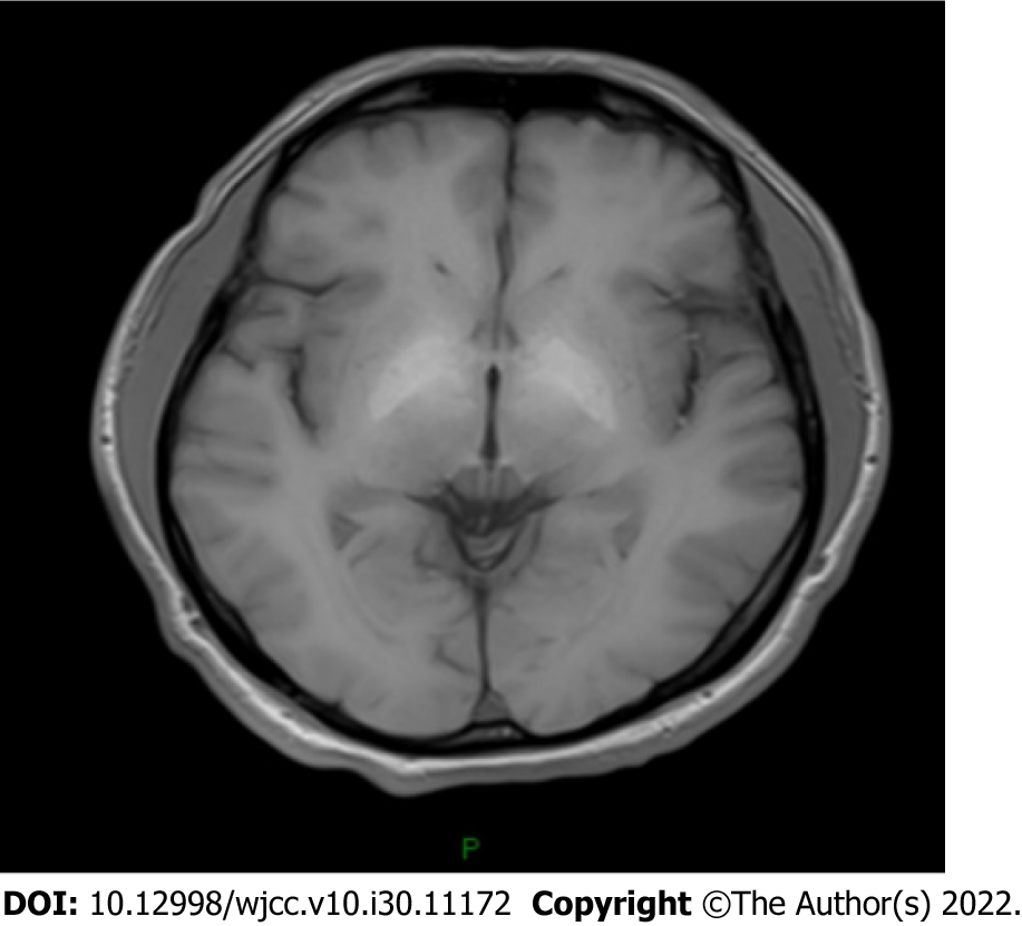
Figure 2 The cranial magnetic resonance imaging revealed increased T1W symmetric signal in the bilateral globus pallidus.
- Citation: Chang CY, Liu C, Duan FF, Zhai H, Song SS, Yang S. Spontaneous remission of hepatic myelopathy in a patient with alcoholic cirrhosis: A case report. World J Clin Cases 2022; 10(30): 11172-11177
- URL: https://www.wjgnet.com/2307-8960/full/v10/i30/11172.htm
- DOI: https://dx.doi.org/10.12998/wjcc.v10.i30.11172


