Copyright
©The Author(s) 2022.
World J Clin Cases. Jan 21, 2022; 10(3): 1056-1066
Published online Jan 21, 2022. doi: 10.12998/wjcc.v10.i3.1056
Published online Jan 21, 2022. doi: 10.12998/wjcc.v10.i3.1056
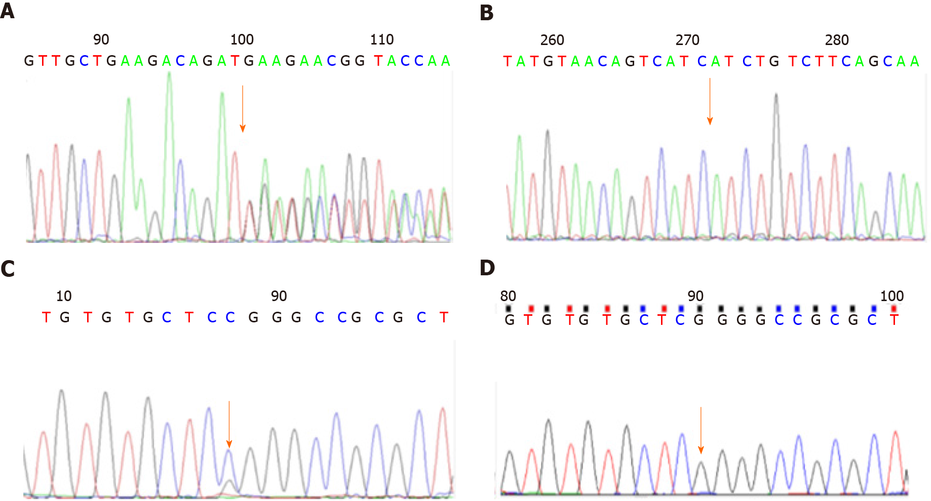
Figure 1 Gene detection results for case 1.
A: c.1068dup T (p.D357_D358delin sX) mutant type; B: c.1068dup T (p.D357_D358delin sX) wild type; C: c.40G>C (p.G14R) mutant type; D: c.40G>C (p.G14R) wild type.
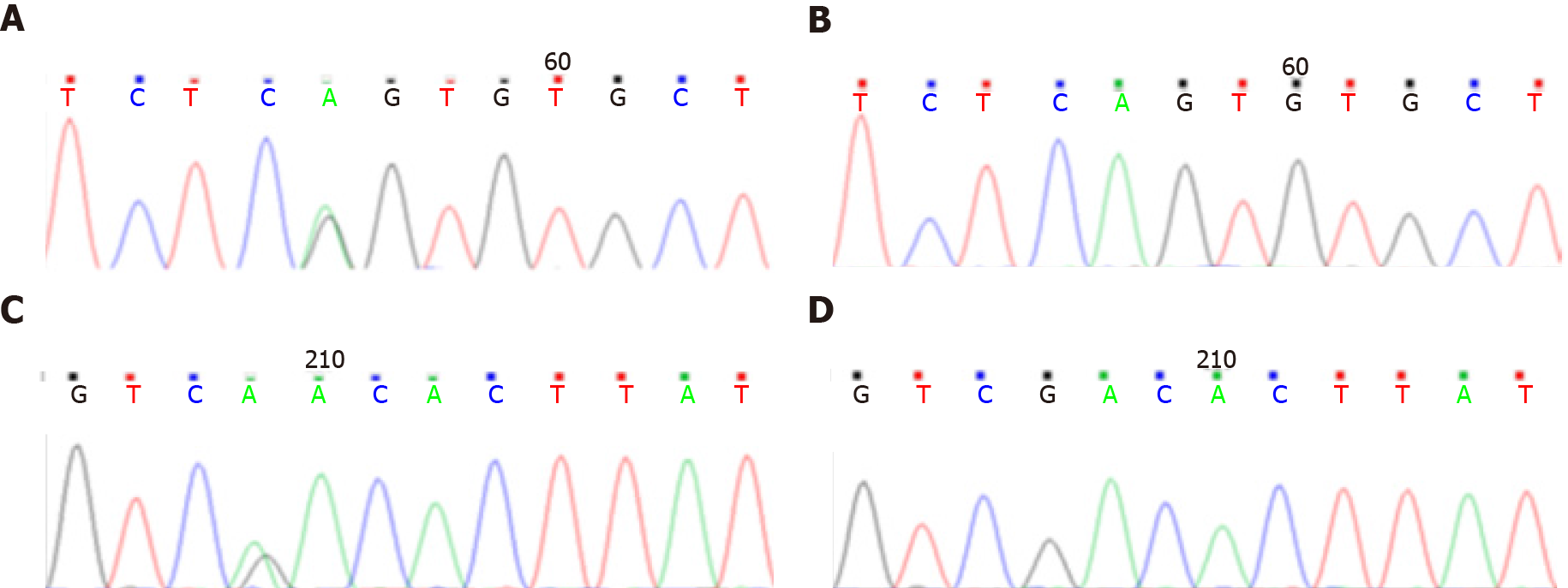
Figure 2 Gene detection results for case 2.
A: c.261-2A>G mutant type; B: c.261-2A>G wild type; C: c.979G>A (p.D327N) mutant type; D: c.979G>A (p.D327N) wild type.
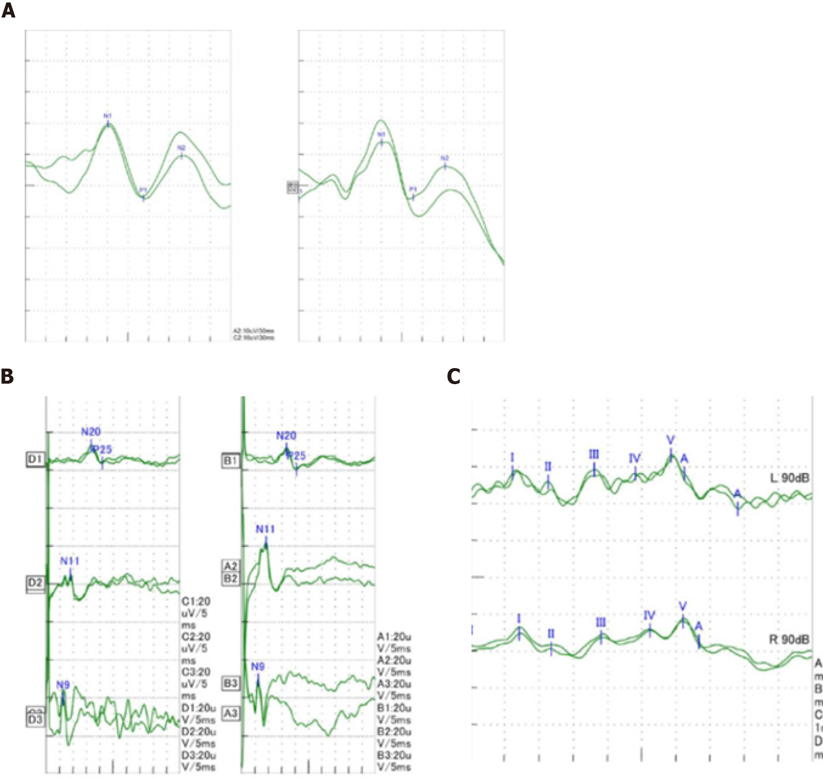
Figure 3 Visual and auditory evoked potentials and somatosensory evoked potentials of the child at 2 years and 3 mo of age.
A: Visual evoked potentials of the child; B: Somatosensory evoked potentials of the child; C: Auditory evoked potentials of the child.
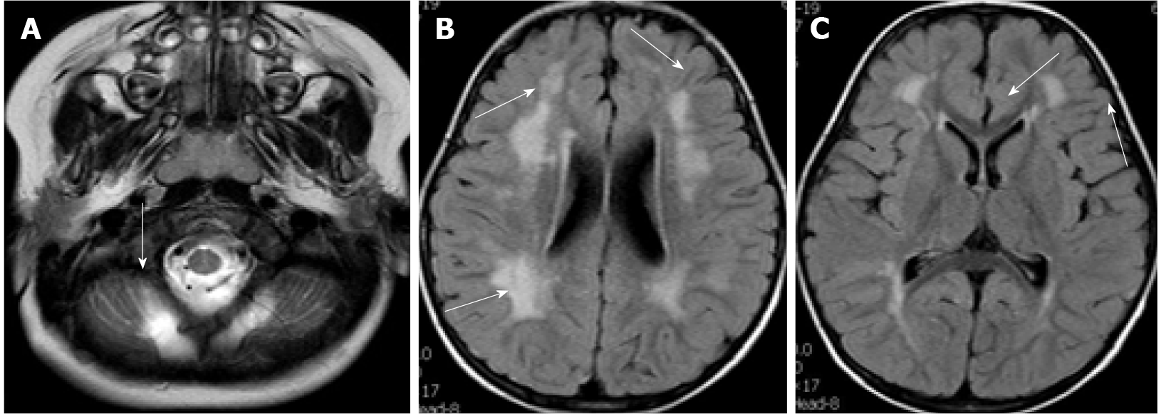
Figure 4 Head magnetic resonance imaging of case 1 at 2 years and 3 mo of age.
A: Cerebellar cyst; B and C: Multiple flaky high FLAIR signals around the bilateral ventricles.
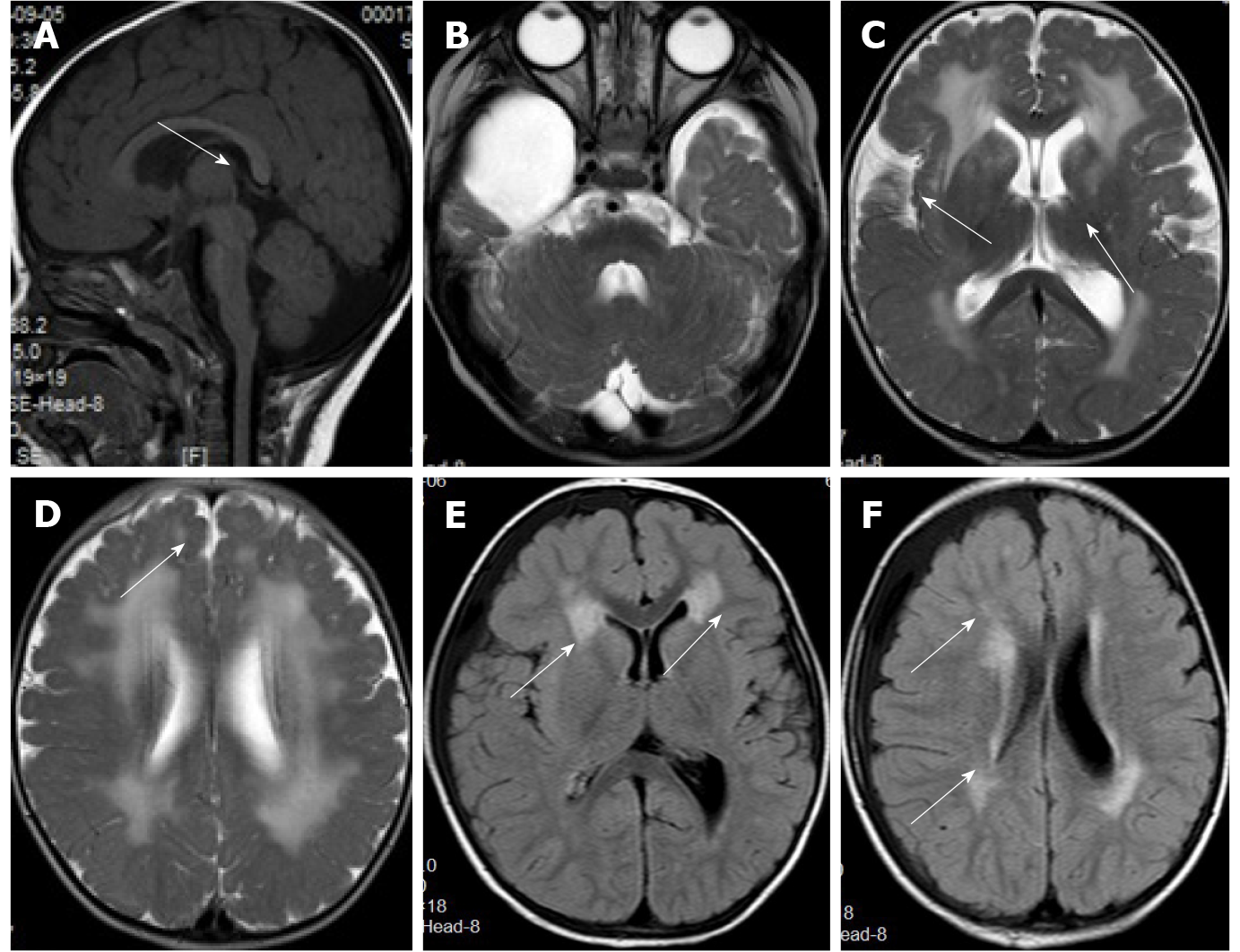
Figure 5 Head magnetic resonance imaging of case 2 at 10 mo and at 2 years and 11 mo of age.
A: Flat pons.; B: Arachnoid cyst in the right temporal pole; C and D: Abnormal signal of periventricular white matter neuronal migration disorder in bilateral frontal lobes; E and F: An abnormal signal of periventricular white matter, which was absorbed and considerably improved compared to that seen in the previous magnetic resonance imaging.
- Citation: Wu WJ, Sun SZ, Li BG. Congenital muscular dystrophy caused by beta1,3-N-acetylgalactosaminyltransferase 2 gene mutation: Two case reports. World J Clin Cases 2022; 10(3): 1056-1066
- URL: https://www.wjgnet.com/2307-8960/full/v10/i3/1056.htm
- DOI: https://dx.doi.org/10.12998/wjcc.v10.i3.1056


