Copyright
©The Author(s) 2022.
World J Clin Cases. Aug 16, 2022; 10(23): 8249-8254
Published online Aug 16, 2022. doi: 10.12998/wjcc.v10.i23.8249
Published online Aug 16, 2022. doi: 10.12998/wjcc.v10.i23.8249
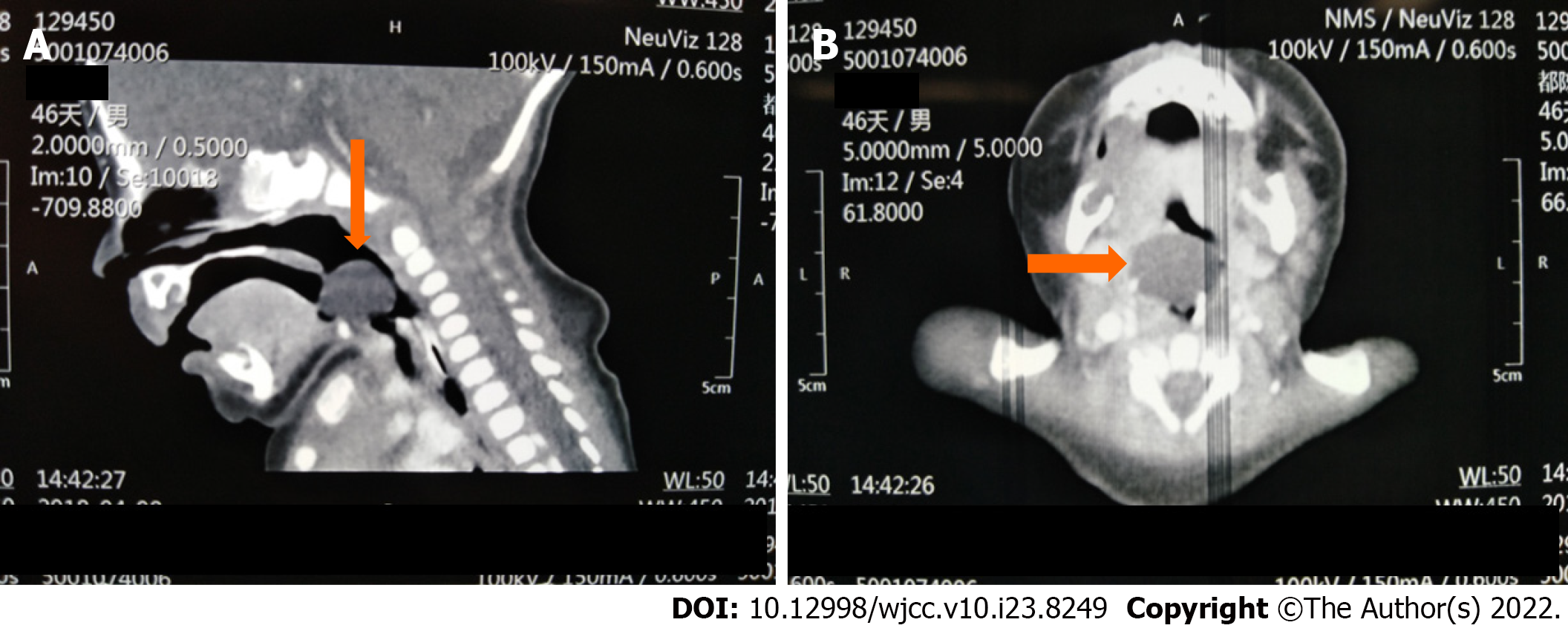
Figure 1 The preoperative computed tomography imaging of head showing a giant round mass on the lingual surface of the epiglottis.
A: Sagittal computed tomography (CT) scan (The yellow arrow indicates the epiglottic cyst); B: Transverse CT scan (The yellow arrow indicates the epiglottic cyst).
Figure 2 Video laryngoscopy confirmed the giant cyst to be implanted on the tip of the epiglottis.
Aspiration was used by an 18-gauge needle.
Figure 3 Video laryngoscopy imaging after epiglottic cysts aspiration.
Epiglottis and arytenoid cartilage appeared.
- Citation: Zheng JQ, Du L, Zhang WY. Aspiration as the first-choice procedure for airway management in an infant with large epiglottic cysts: A case report. World J Clin Cases 2022; 10(23): 8249-8254
- URL: https://www.wjgnet.com/2307-8960/full/v10/i23/8249.htm
- DOI: https://dx.doi.org/10.12998/wjcc.v10.i23.8249


