Copyright
©The Author(s) 2022.
World J Clin Cases. Jul 6, 2022; 10(19): 6716-6721
Published online Jul 6, 2022. doi: 10.12998/wjcc.v10.i19.6716
Published online Jul 6, 2022. doi: 10.12998/wjcc.v10.i19.6716
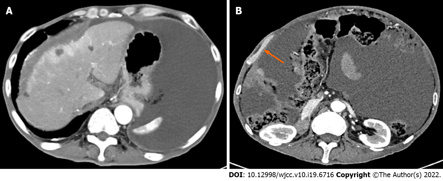
Figure 1 Computed tomography images.
A: Abdominal computed tomography revealed multiple hepatic cysts, liver cirrhosis and ascites; B: The right peritoneum was irregularly thickened.
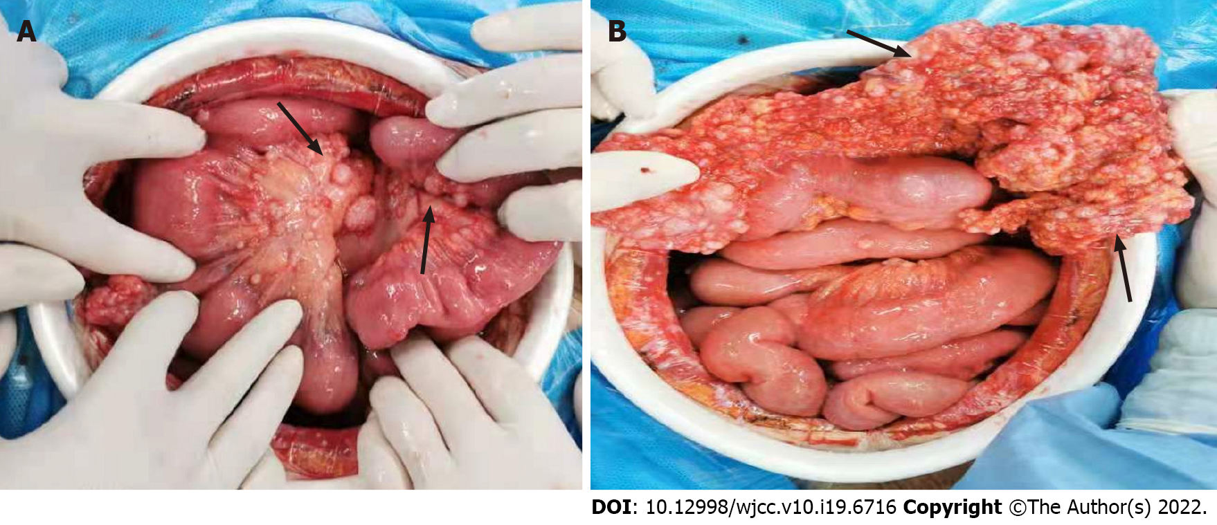
Figure 2
The greater omentum was pancake-shaped, multiple round masses were observed in the parietal and visceral peritoneum, with a diameter of 3-20 mm and the right subphrenic peritoneum was thickened obviously, with an area of about 15 cm × 15 cm.
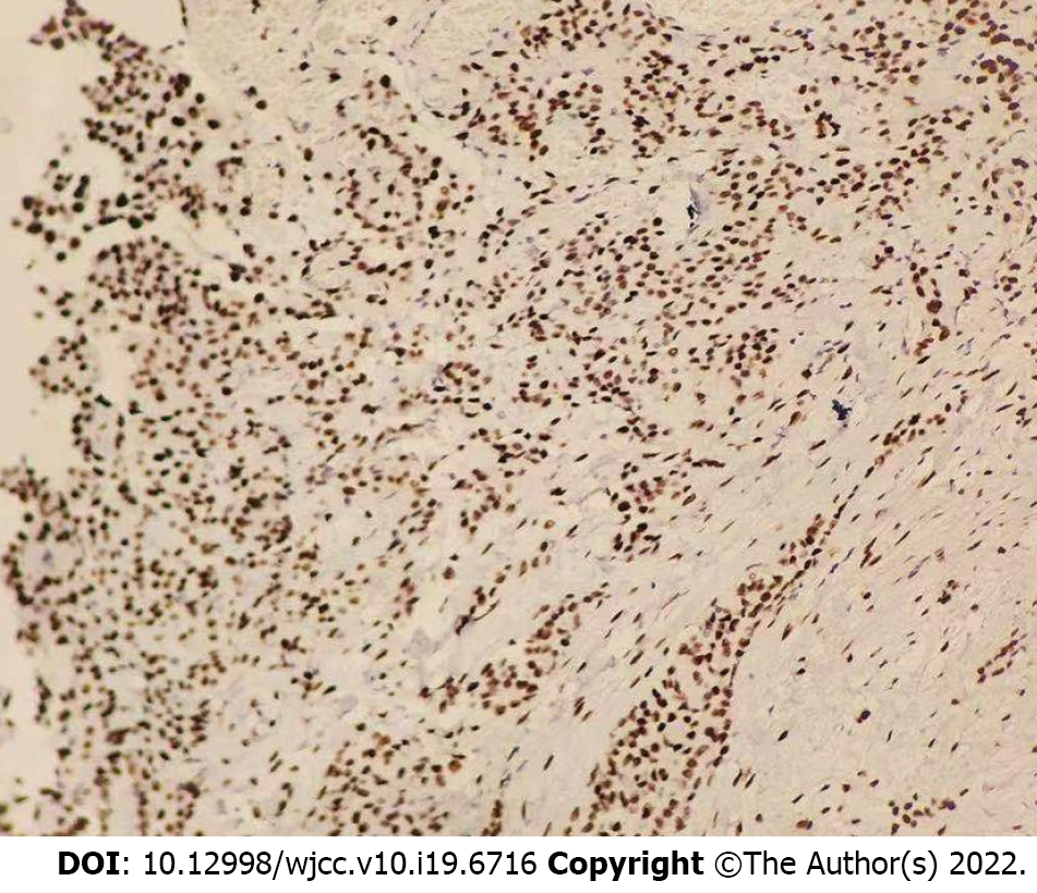
Figure 3
Pathological and immunohistochemical examinations showed an epithelioid malignant peritoneal mesothelioma.
- Citation: Liu L, Zhu XY, Zong WJ, Chu CL, Zhu JY, Shen XJ. Concurrent alcoholic cirrhosis and malignant peritoneal mesothelioma in a patient: A case report. World J Clin Cases 2022; 10(19): 6716-6721
- URL: https://www.wjgnet.com/2307-8960/full/v10/i19/6716.htm
- DOI: https://dx.doi.org/10.12998/wjcc.v10.i19.6716


