Copyright
©The Author(s) 2022.
World J Clin Cases. Jul 6, 2022; 10(19): 6688-6694
Published online Jul 6, 2022. doi: 10.12998/wjcc.v10.i19.6688
Published online Jul 6, 2022. doi: 10.12998/wjcc.v10.i19.6688
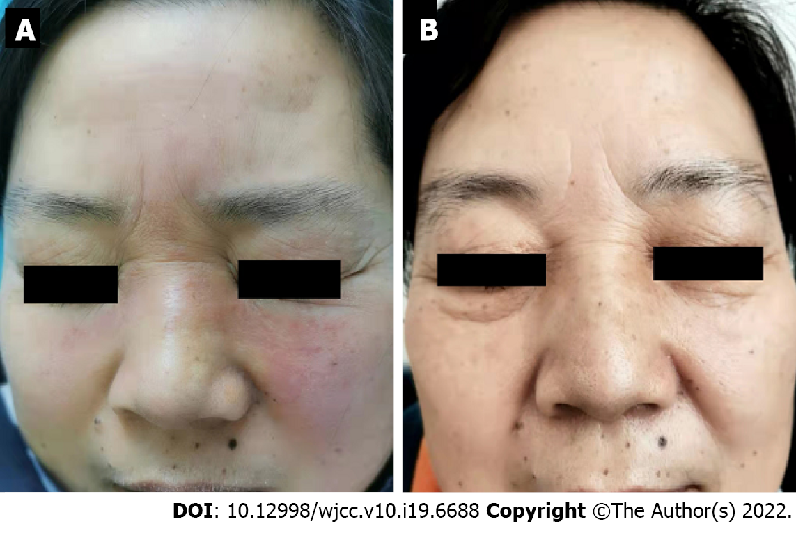
Figure 1 Facial lesions of the patient with Morbihan disease.
A: Non-pitting edema and erythema of the upper two-thirds of the face; B: The lesions disappeared completely after 4 mo of treatment with total glucosides of paeony capsules, and follow-up of almost 1 yr showed no recurrence.
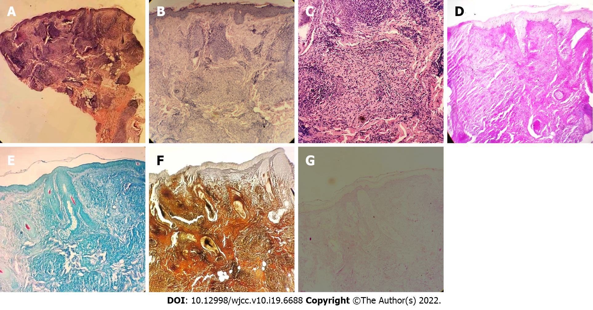
Figure 2 Histopathology of the patient's facial skin lesions.
A-C: Hyperkeratosis of the epidermis, nodular inflammatory lesions in the dermis, epithelioid granuloma, and inflammatory infiltrating cells dominated by lymphocytes and histiocytes around skin appendages and blood vessels (HE staining A: × 4; B: × 20; C: × 40); D: Negative for Alcian blue staining (× 20); E: Negative for acid fast staining (× 20); F: Negative for silver staining (× 20); G: Negative for periodic acid-Schiff staining (× 20).
- Citation: Zhou LF, Lu R. Successful treatment of Morbihan disease with total glucosides of paeony: A case report. World J Clin Cases 2022; 10(19): 6688-6694
- URL: https://www.wjgnet.com/2307-8960/full/v10/i19/6688.htm
- DOI: https://dx.doi.org/10.12998/wjcc.v10.i19.6688


