Copyright
©The Author(s) 2017.
World J Transplant. Aug 24, 2017; 7(4): 243-249
Published online Aug 24, 2017. doi: 10.5500/wjt.v7.i4.243
Published online Aug 24, 2017. doi: 10.5500/wjt.v7.i4.243
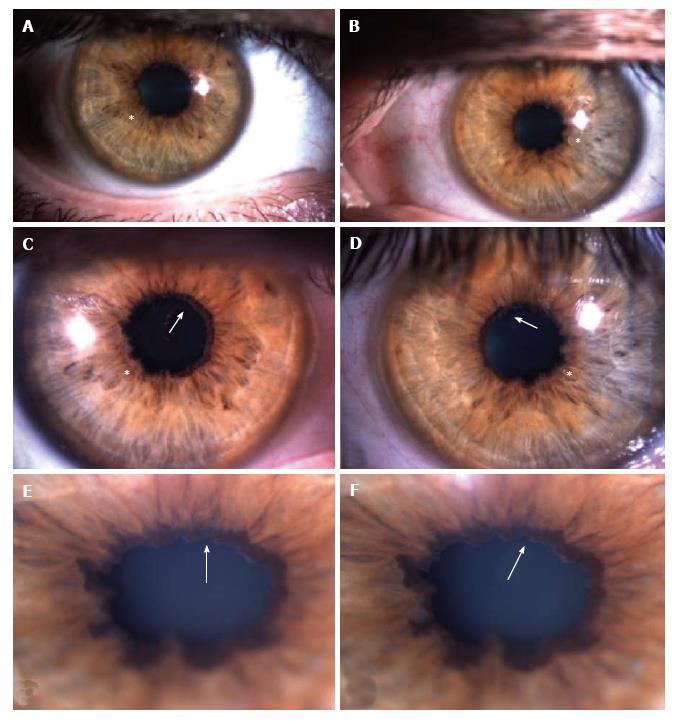
Figure 1 Slit-lamp photos showing scalloped pupils (astherisks) and amyloid deposition in the pupillary border (arrows) in both eyes.
A and B: Slit-lamp photos of anterior segment of right (A) and left eyes (B) at low magnification; C and D: Slit-lamp photos at higher magnification to show pupillary margins of right (C) and left (D) eyes with more detail, in order to highlight the irregular pupillary margins, the scalloped pupils (astherisks) with amyloid deposits (arrows); E and F: Slit-lamp photos of the right eye (E and F) at the highest magnification to enhance visualization of the pupillary amyloid deposits (arrows), which resemble those seen in pseudoexfoliation syndrome.
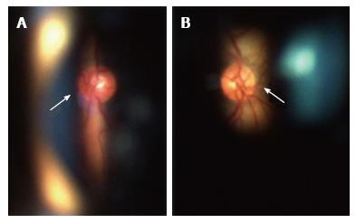
Figure 2 Fundoscopy of right (A) and left (B) eyes showed normal-appearing optic discs (arrows) and absence of abnormalities in posterior pole and peripheral retina.
Ocular fundus was perfectly visible due to mild amyloid deposition in the vitreous.
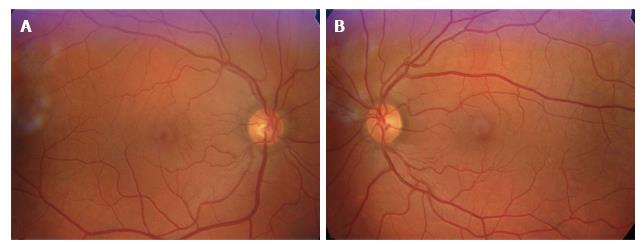
Figure 3 Retinography of right eye (A) and left eye (B) showed normal posterior poles, which were clearly visible due to the mild amyloid deposition in the vitreous, with only few opacities, which did not compromise visual acuity.
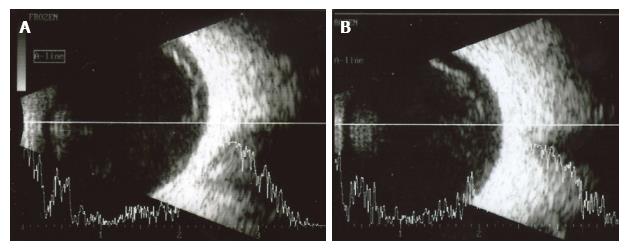
Figure 4 Ophthalmic ultrasound of right (A) and left (B) eyes showing some vitreous opacities corresponding to amyloid deposits in the vitreous.
This amyloid deposition in the vitreous is relatively mild compared to the degree of amyloid deposition in anterior segment.
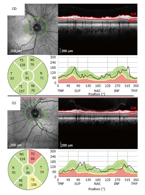
Figure 5 Optical coherence tomography showed a localized defect in the temporal-superior area of the peripapillary retinal nerve fiber layer of the left eye (OS).
In the right eye (OD), the retinal nerve fiber layer thickness was normal in all peripapillary locations.
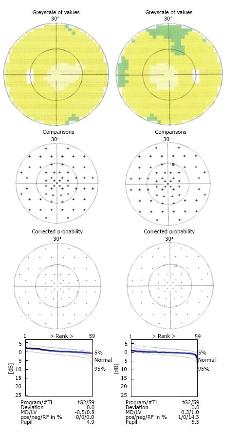
Figure 6 Computerized static perimetry of right eye (at the right) and left eye (at the left) (tendency-oriented perimetry, TOP - 30º program, Octopus 101 perimeter, Haag-Streit Diagnostics, Switzerland) in November 2011, showed the absence of clinically significant abnormalities in the visual fields - preperimetic glaucoma.
- Citation: Gama IF, Almeida LD. De novo intraocular amyloid deposition after hepatic transplantation in familial amyloidotic polyneuropathy. World J Transplant 2017; 7(4): 243-249
- URL: https://www.wjgnet.com/2220-3230/full/v7/i4/243.htm
- DOI: https://dx.doi.org/10.5500/wjt.v7.i4.243


