Copyright
©The Author(s) 2017.
World J Transplant. Aug 24, 2017; 7(4): 243-249
Published online Aug 24, 2017. doi: 10.5500/wjt.v7.i4.243
Published online Aug 24, 2017. doi: 10.5500/wjt.v7.i4.243
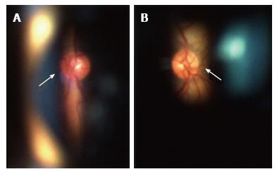
Figure 2 Fundoscopy of right (A) and left (B) eyes showed normal-appearing optic discs (arrows) and absence of abnormalities in posterior pole and peripheral retina.
Ocular fundus was perfectly visible due to mild amyloid deposition in the vitreous.
- Citation: Gama IF, Almeida LD. De novo intraocular amyloid deposition after hepatic transplantation in familial amyloidotic polyneuropathy. World J Transplant 2017; 7(4): 243-249
- URL: https://www.wjgnet.com/2220-3230/full/v7/i4/243.htm
- DOI: https://dx.doi.org/10.5500/wjt.v7.i4.243


