Copyright
©The Author(s) 2025.
World J Exp Med. Sep 20, 2025; 15(3): 104431
Published online Sep 20, 2025. doi: 10.5493/wjem.v15.i3.104431
Published online Sep 20, 2025. doi: 10.5493/wjem.v15.i3.104431
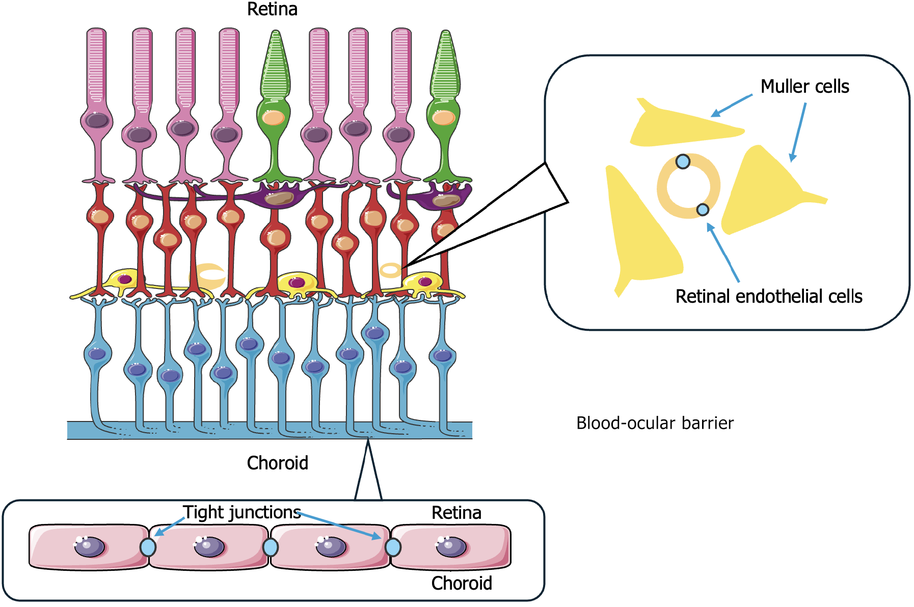
Figure 1 Blood-ocular barrier maintains the immune-privileged status of the eye by restricting the entry of blood-borne substances.
It consists of two main components: The inner blood-retinal barrier formed by tight junctions between retinal endothelial cells and supported by Müller glial cells, which regulate fluid and ion balance and the outer blood-retinal barrier composed of tight junctions between retinal pigment epithelial cells adjacent to the choroid, controlling molecular exchange between the retina and the systemic circulation.
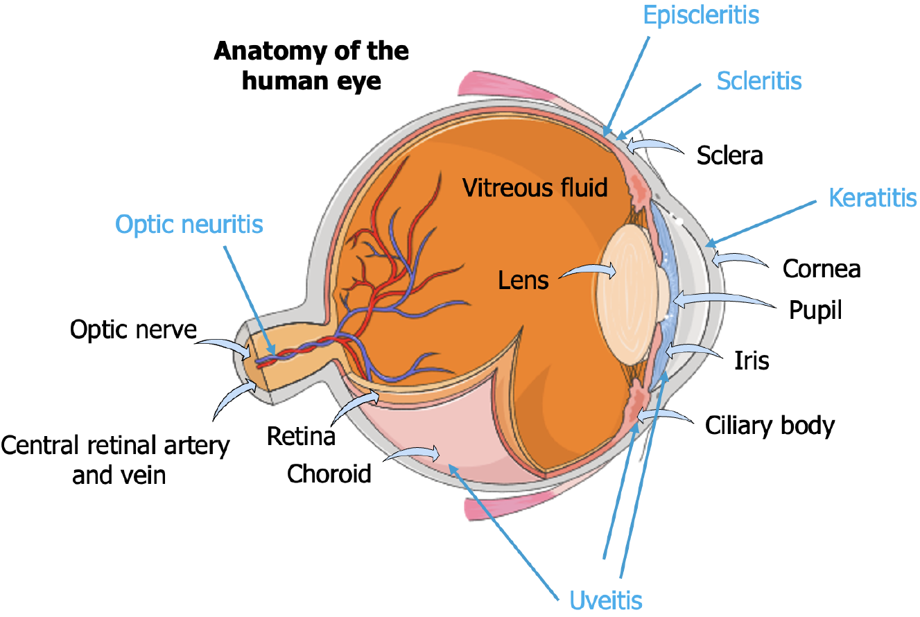
Figure 2 Anatomy of the human eye and locations of the most common manifestations of autoimmune diseases that affect the eye.
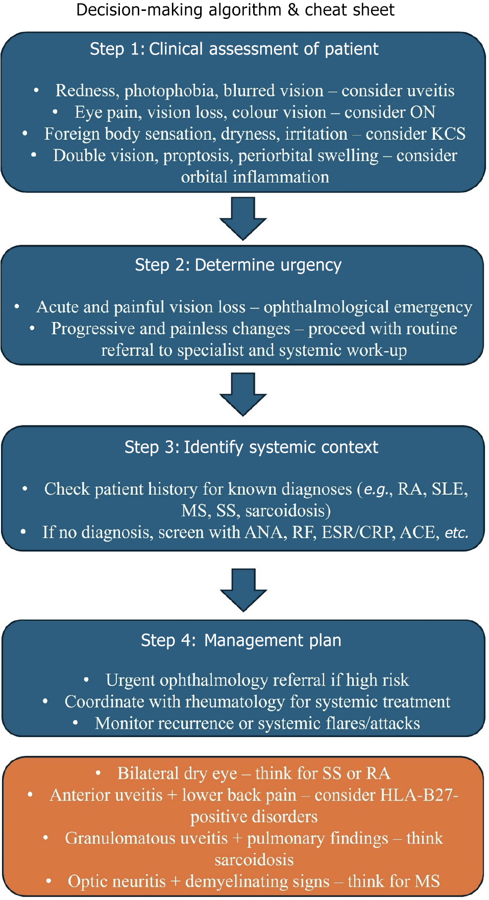
Figure 3 Diagnostic algorithm for systemic work-up in patients with vision loss suspected of an autoimmune etiology.
ON: Optical neuritis; SS: Sjögren's syndrome; RA: Rheumatoid arthritis; MS: Multiple sclerosis; KCS: Keratoconjunctivitis sicca; SLE: Systemic lupus erythematosus; ANA: Anti-nuclear antibodies; RF: Rheumatoid factor; ESR: Erythrocyte sedimentation rate; CRP: C-reactive protein; ACE: Angiotensin converting enzyme; HLA: Human leukocyte antigen.
- Citation: El Kaouri I, Deligiannis I, Bakopoulou K, Sdralis PP, Shoumnalieva-Ivanova V, Shumnalieva R, Velikova T. Ophthalmological manifestations in autoimmune diseases: Overcoming diagnostic and therapeutic challenges. World J Exp Med 2025; 15(3): 104431
- URL: https://www.wjgnet.com/2220-315X/full/v15/i3/104431.htm
- DOI: https://dx.doi.org/10.5493/wjem.v15.i3.104431


