Copyright
©The Author(s) 2025.
World J Clin Pediatr. Sep 9, 2025; 14(3): 102898
Published online Sep 9, 2025. doi: 10.5409/wjcp.v14.i3.102898
Published online Sep 9, 2025. doi: 10.5409/wjcp.v14.i3.102898
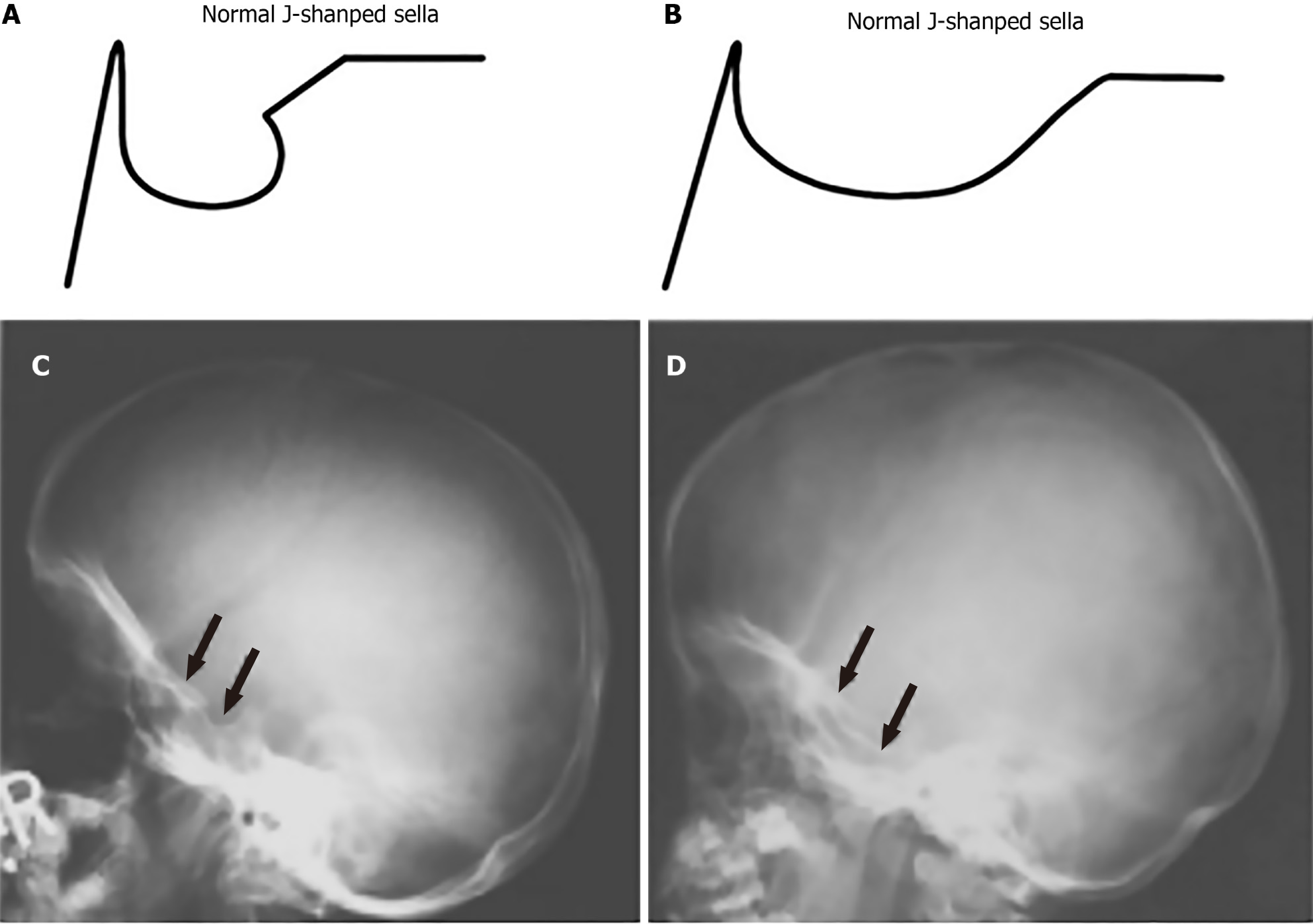
Figure 1 Comparison of normal and abnormal J-shaped sella in lateral skull X-rays.
A: Schematic representation of a normal J-shaped sella; B: An abnormal J-shaped sella with a more pronounced deformation; C: Lateral skull radiograph showing a normal J-shaped sella (arrows); D: Lateral skull radiograph depicting an abnormal J-shaped sella (arrows), frequently seen in patients with mucopolysaccharidoses.
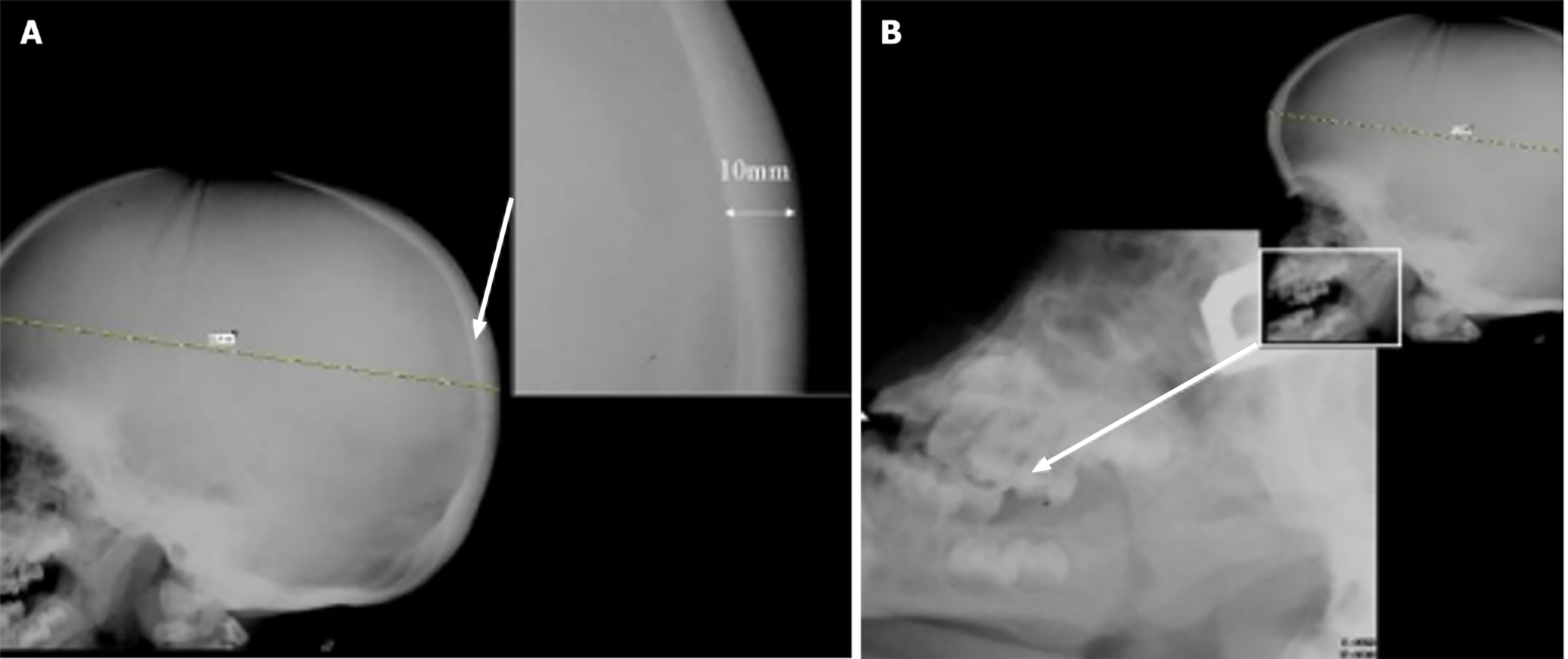
Figure 2 Lateral skull X-ray.
A: Showing marked thickening of the calvarium (10 mm) in the posterior two-thirds of the skull vault, a radiographic feature frequently observed in mucopolysaccharidoses. The inset provides a magnified view to emphasize the increased thickness; B: Demonstrating progenia (enlarged mandible) due to the enlarged mandible (arrow) and square mandibular condyles are present.
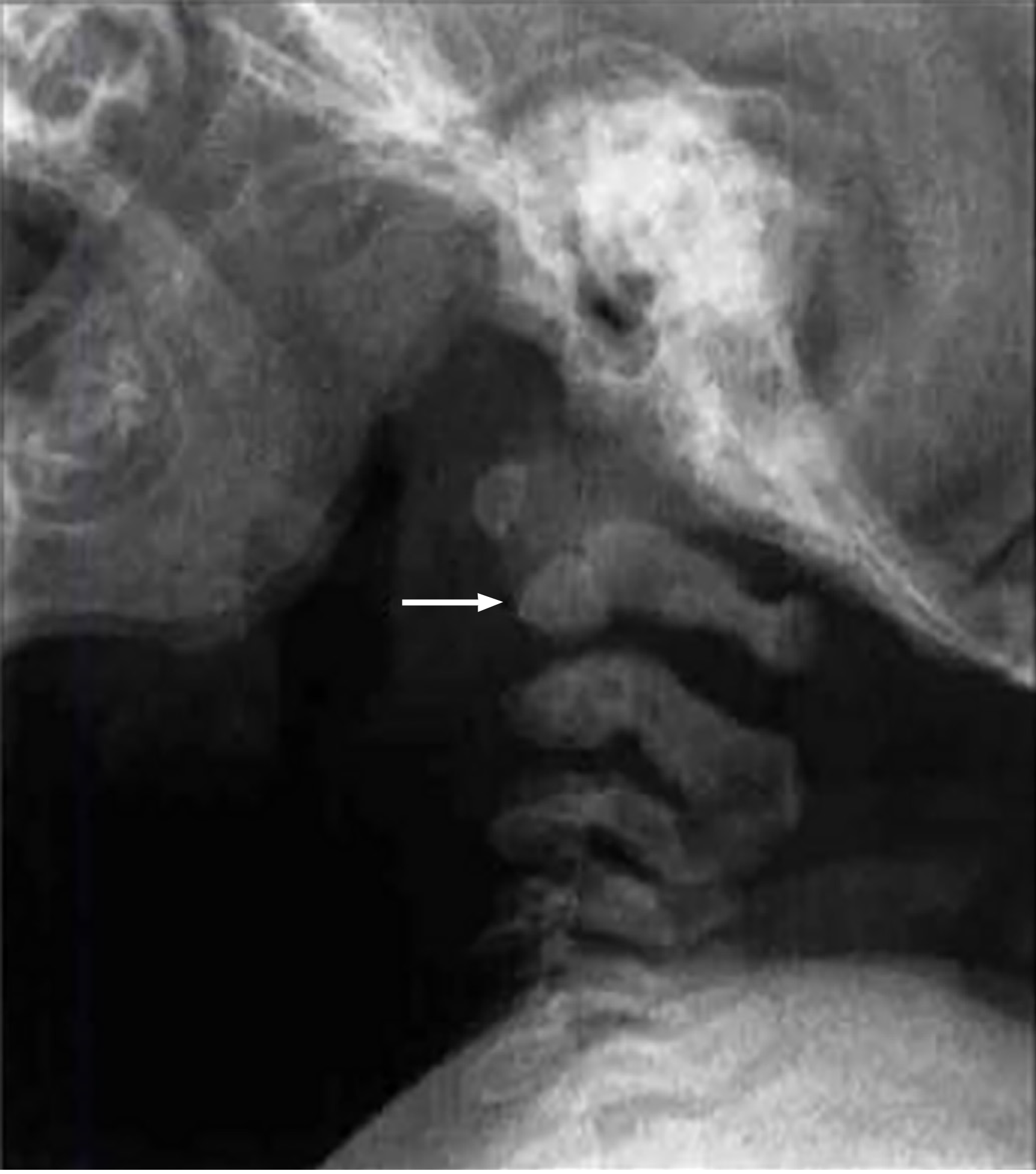
Figure 3 Lateral radiograph shows hypoplastic odontoid process (arrow).
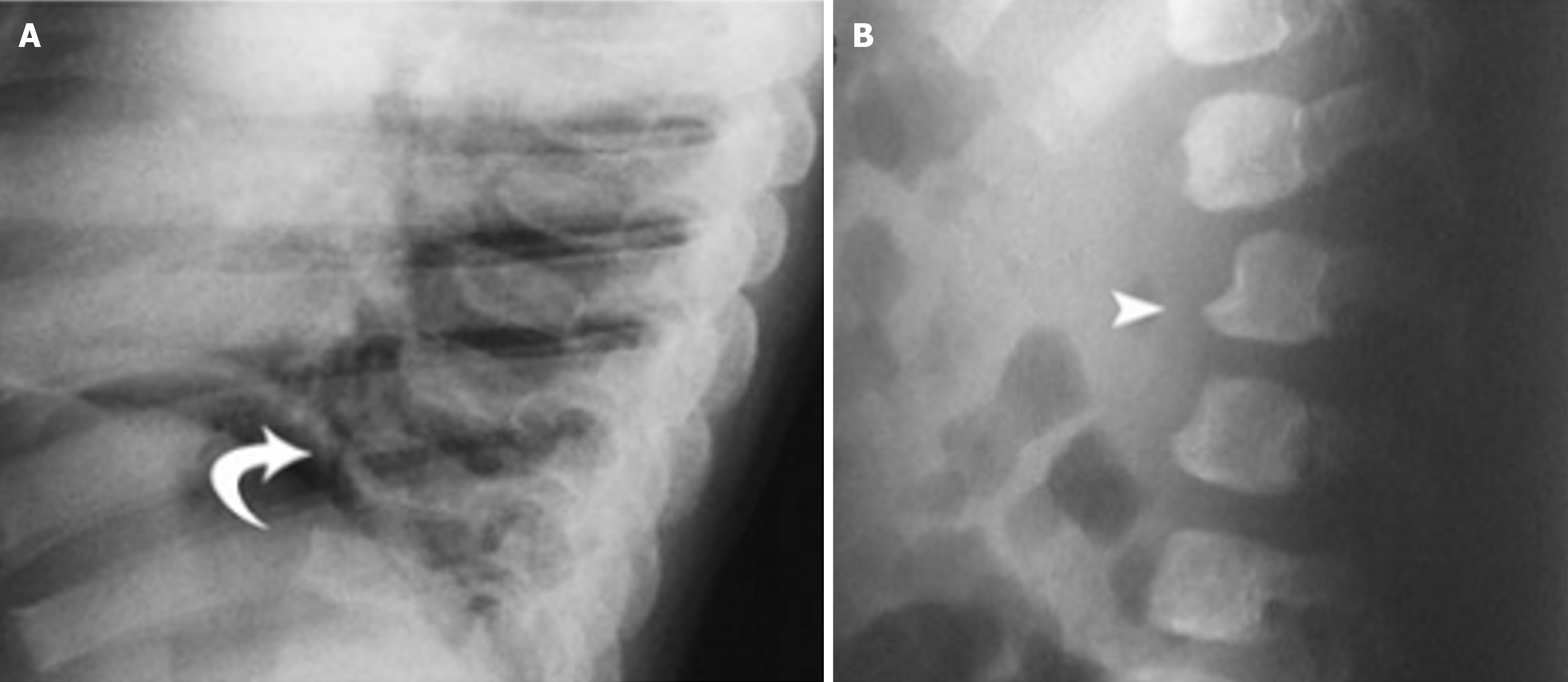
Figure 4 Image on the left showing.
A: “Anterior beaking” with posterior scalloping due to deficient anterosuperior corner; B: Platyspondylia (flat vertebral bodies) with “wedge-shaped” deformity.
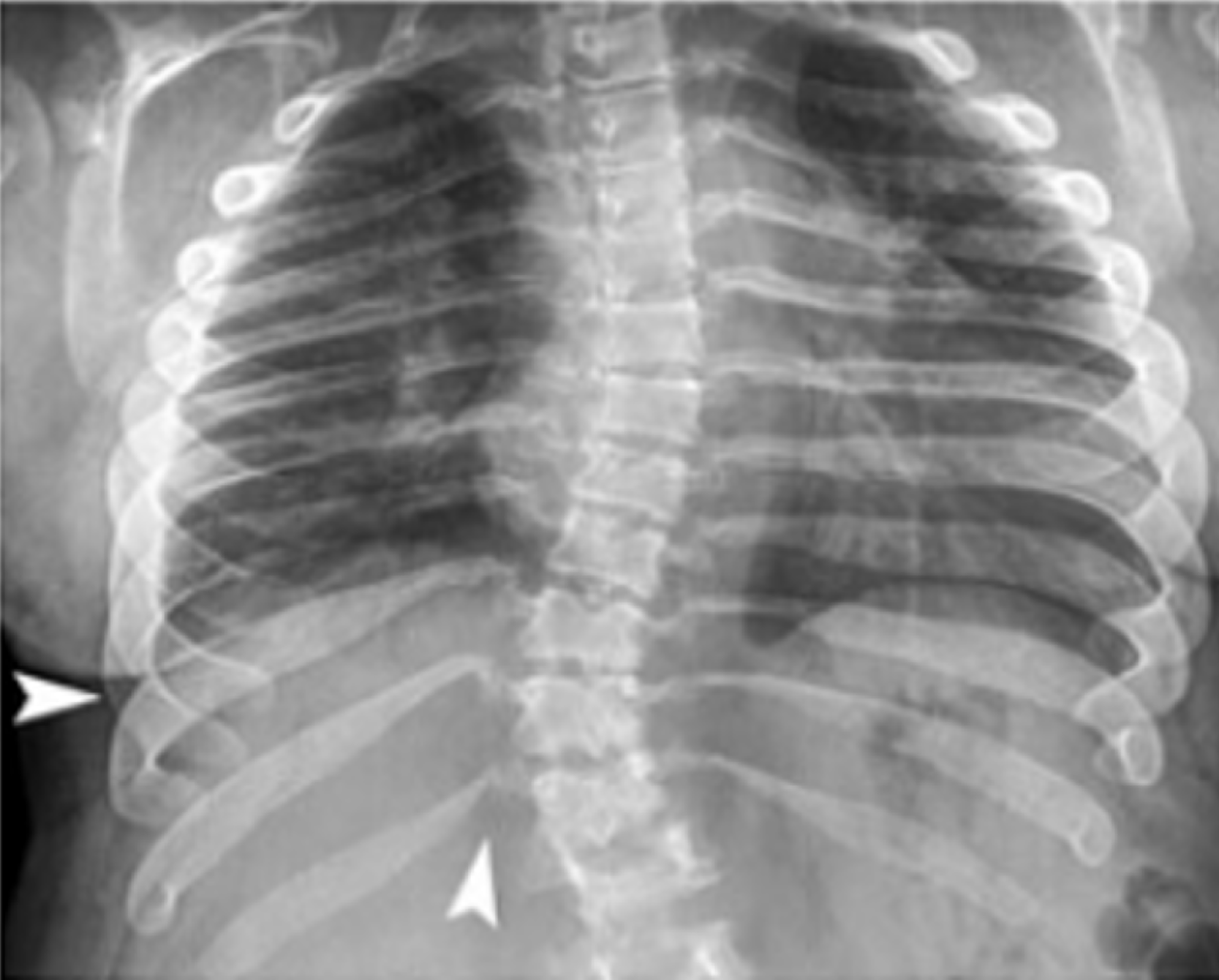
Figure 5 Paddle or oar shaped ribs (white arrowheads) tapered proximally and wider distally.
Figure 6 Broad and short clavicles (arrow).
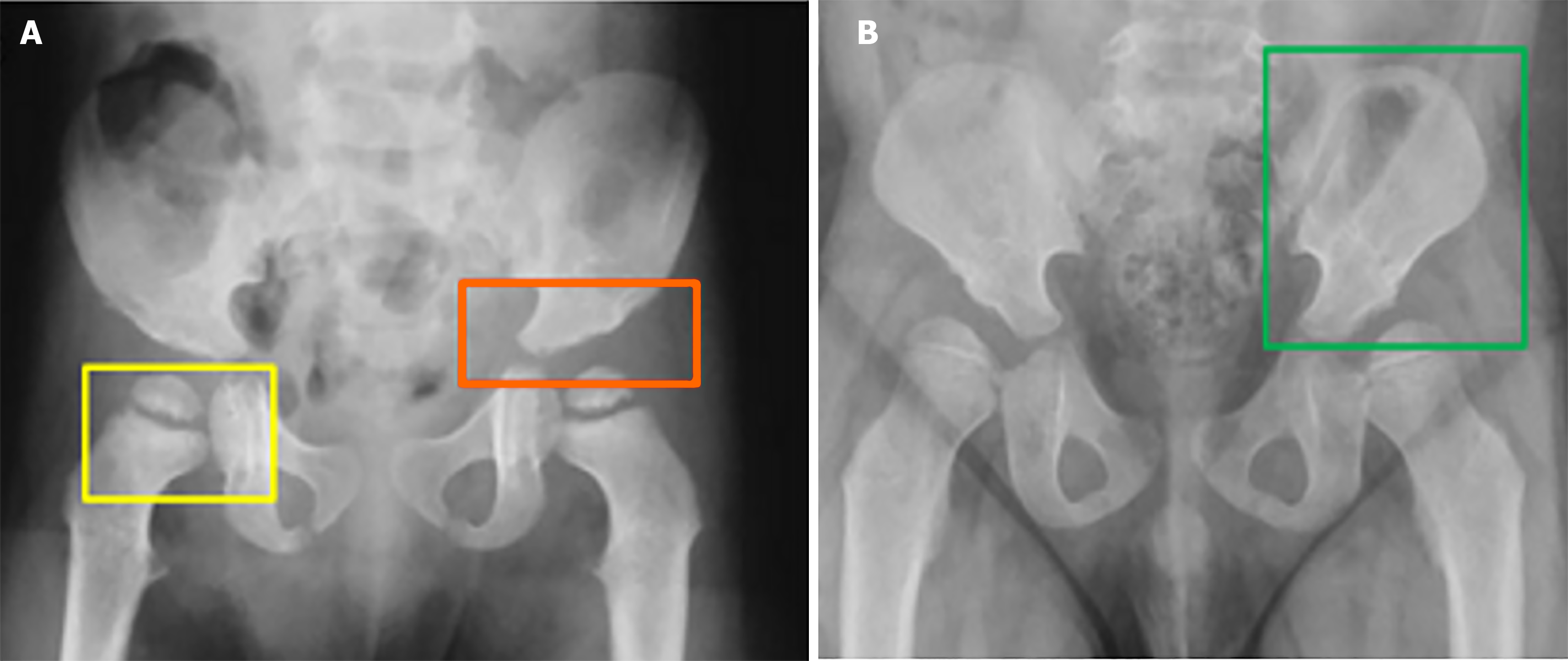
Figure 7 Patient pelvic condition.
A: Images illustrating a poorly formed acetabulum (yellow), inferior tapering of the ilea with a poorly developed acetabulum (orange); B: Rounded iliac wings (green).
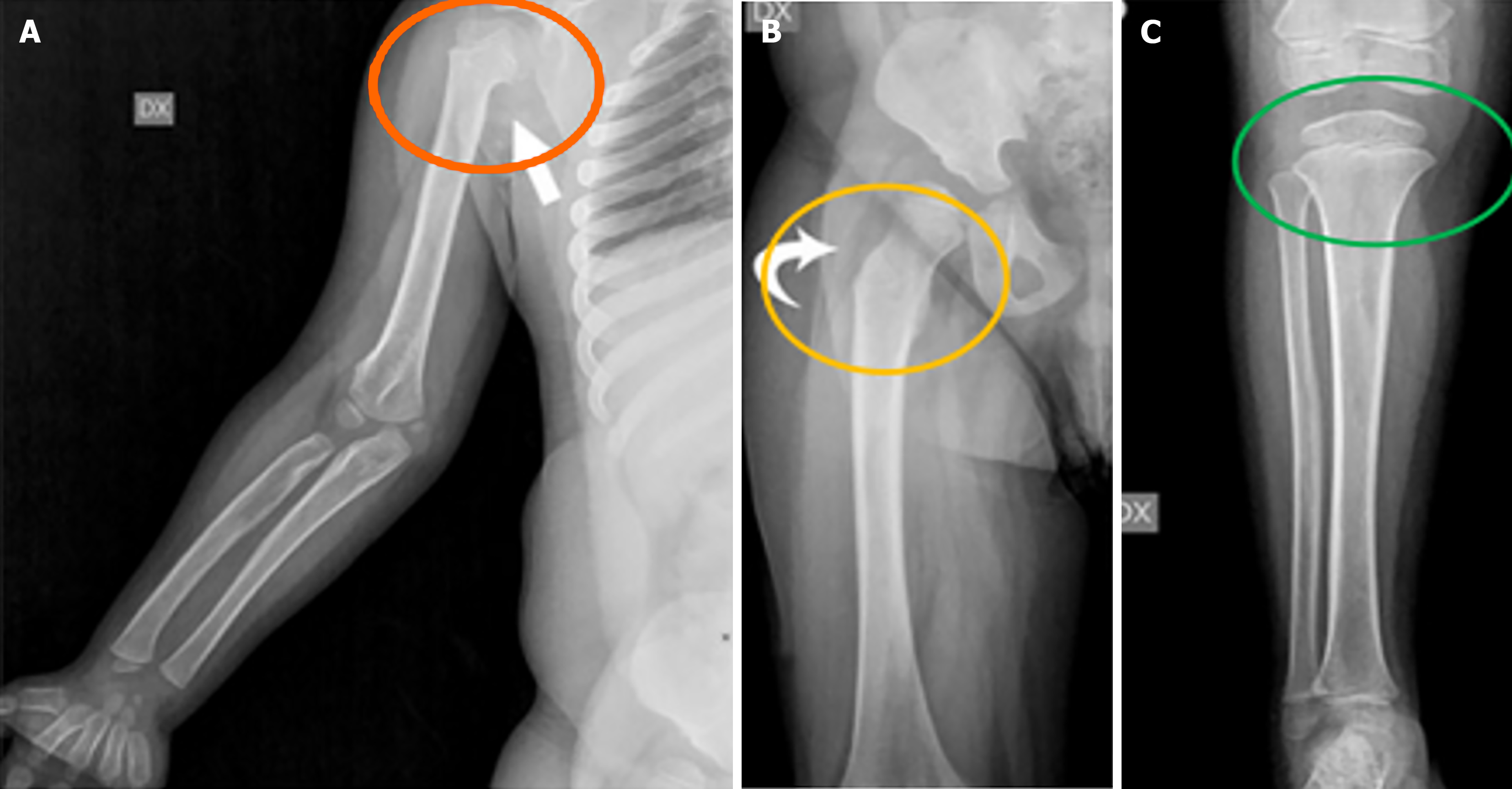
Figure 8 Other bone characteristics of the patient.
A: Proximal humeral notching; B: Long and narrow femoral neck along with an underdeveloped medial portion of the proximal femoral epiphysis; C: Frayed and flared tibial metaphyses.
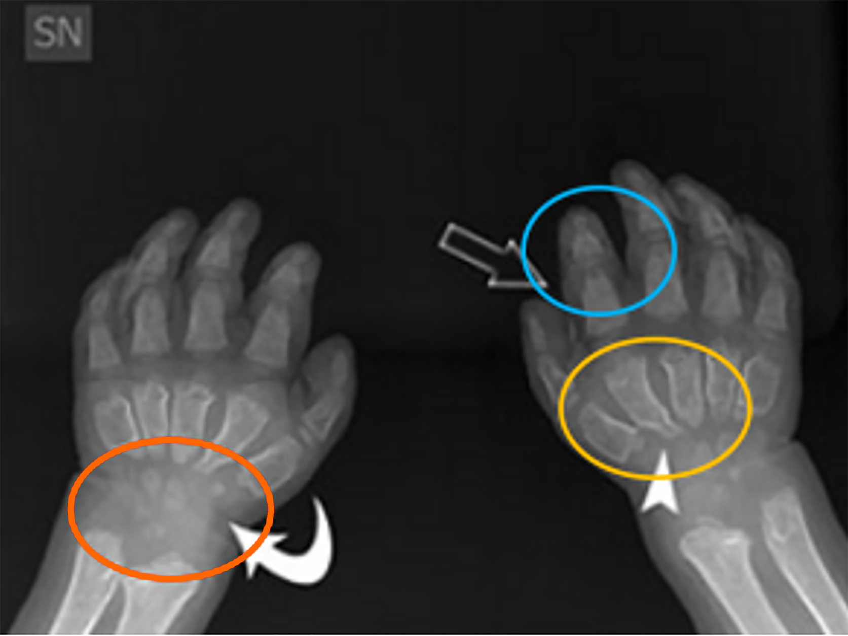
Figure 9 Radiograph showing bullet-shaped phalanges (blue), broad and proximally pointed short metacarpals (yellow) and small irregular carpal bones (orange).
- Citation: Teixeira de Castro Gonçalves Ortega AC, Amorim Moreira Alves G, Perera Molligoda Arachchige AS. Radiographic assessment of mucopolysaccharidoses: A pictorial review. World J Clin Pediatr 2025; 14(3): 102898
- URL: https://www.wjgnet.com/2219-2808/full/v14/i3/102898.htm
- DOI: https://dx.doi.org/10.5409/wjcp.v14.i3.102898


