Copyright
©The Author(s) 2025.
World J Clin Pediatr. Sep 9, 2025; 14(3): 102898
Published online Sep 9, 2025. doi: 10.5409/wjcp.v14.i3.102898
Published online Sep 9, 2025. doi: 10.5409/wjcp.v14.i3.102898
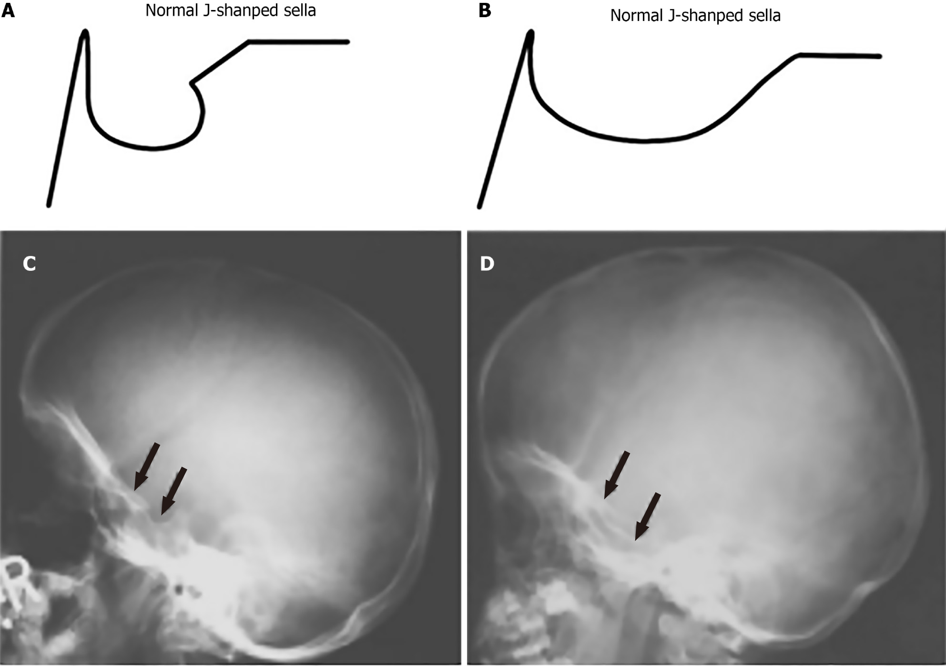
Figure 1 Comparison of normal and abnormal J-shaped sella in lateral skull X-rays.
A: Schematic representation of a normal J-shaped sella; B: An abnormal J-shaped sella with a more pronounced deformation; C: Lateral skull radiograph showing a normal J-shaped sella (arrows); D: Lateral skull radiograph depicting an abnormal J-shaped sella (arrows), frequently seen in patients with mucopolysaccharidoses.
- Citation: Teixeira de Castro Gonçalves Ortega AC, Amorim Moreira Alves G, Perera Molligoda Arachchige AS. Radiographic assessment of mucopolysaccharidoses: A pictorial review. World J Clin Pediatr 2025; 14(3): 102898
- URL: https://www.wjgnet.com/2219-2808/full/v14/i3/102898.htm
- DOI: https://dx.doi.org/10.5409/wjcp.v14.i3.102898


