Copyright
©The Author(s) 2025.
World J Clin Pediatr. Sep 9, 2025; 14(3): 102898
Published online Sep 9, 2025. doi: 10.5409/wjcp.v14.i3.102898
Published online Sep 9, 2025. doi: 10.5409/wjcp.v14.i3.102898
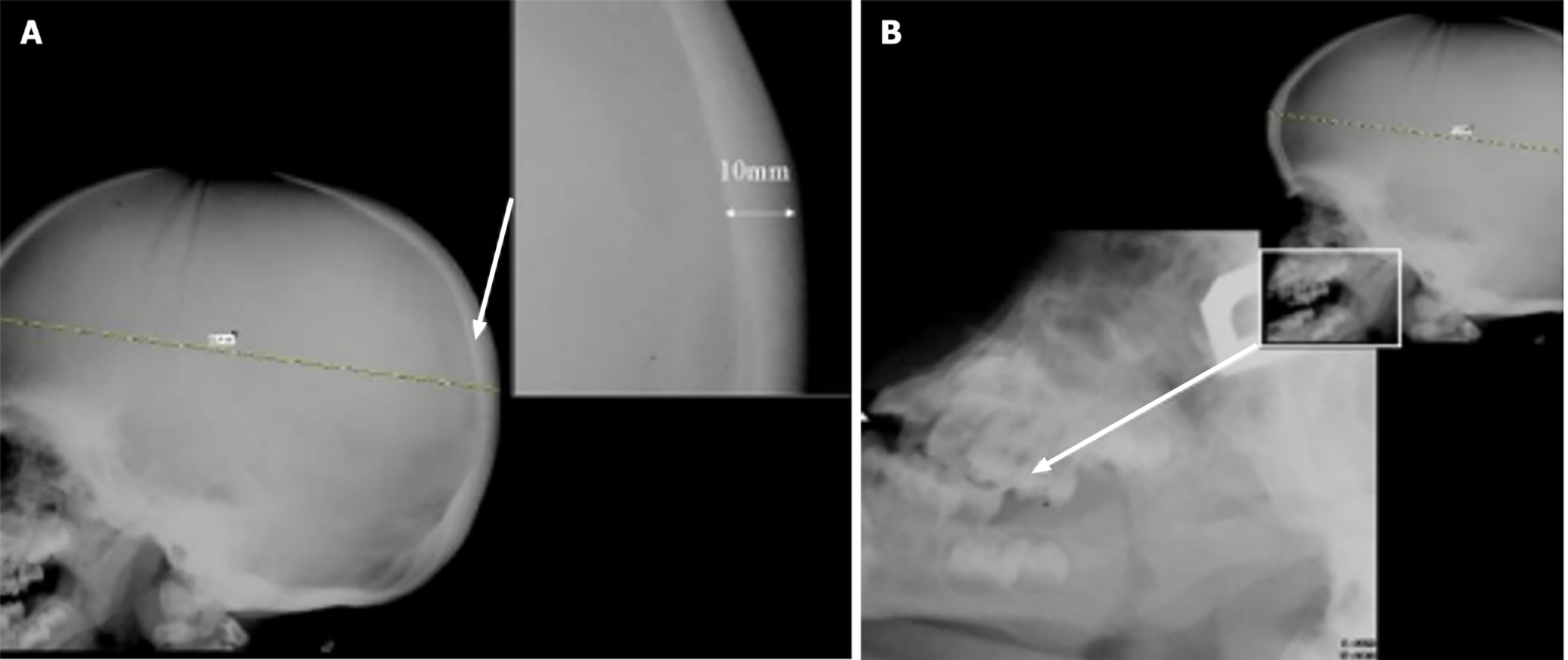
Figure 2 Lateral skull X-ray.
A: Showing marked thickening of the calvarium (10 mm) in the posterior two-thirds of the skull vault, a radiographic feature frequently observed in mucopolysaccharidoses. The inset provides a magnified view to emphasize the increased thickness; B: Demonstrating progenia (enlarged mandible) due to the enlarged mandible (arrow) and square mandibular condyles are present.
- Citation: Teixeira de Castro Gonçalves Ortega AC, Amorim Moreira Alves G, Perera Molligoda Arachchige AS. Radiographic assessment of mucopolysaccharidoses: A pictorial review. World J Clin Pediatr 2025; 14(3): 102898
- URL: https://www.wjgnet.com/2219-2808/full/v14/i3/102898.htm
- DOI: https://dx.doi.org/10.5409/wjcp.v14.i3.102898


