Copyright
©The Author(s) 2025.
World J Clin Pediatr. Sep 9, 2025; 14(3): 102741
Published online Sep 9, 2025. doi: 10.5409/wjcp.v14.i3.102741
Published online Sep 9, 2025. doi: 10.5409/wjcp.v14.i3.102741
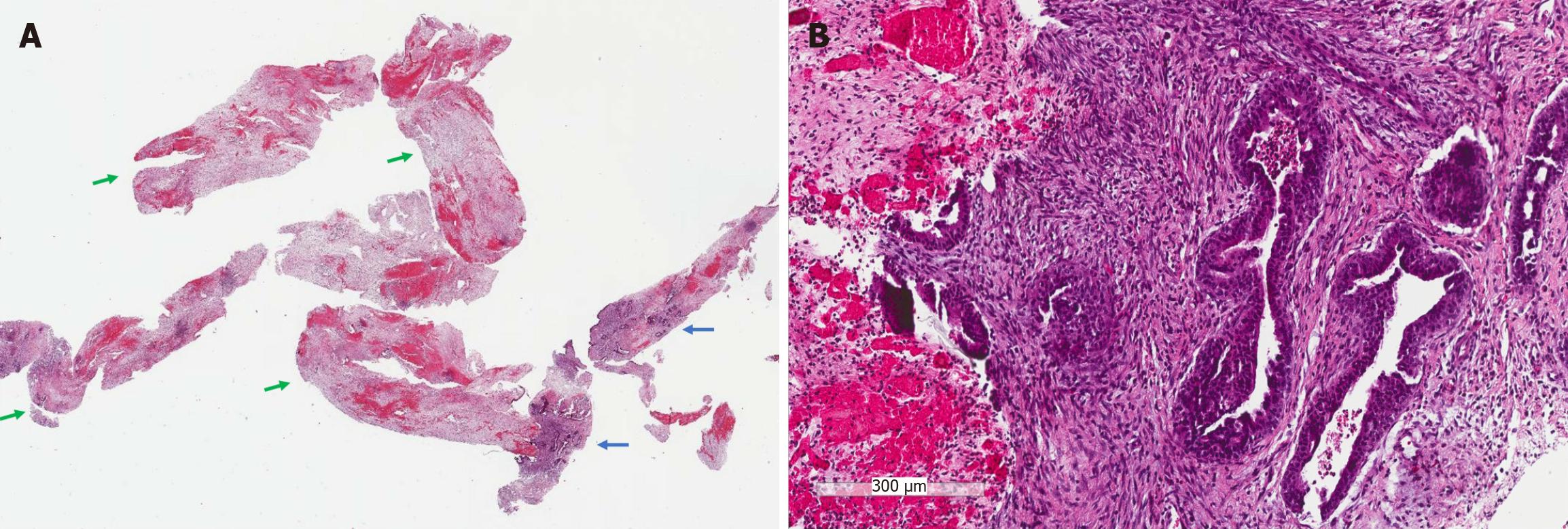
Figure 1 Breast biopsy.
A: The whole slide view showed tissue cores composed predominantly of infarcted tissue (green arrows) admixed with scant viable breast tissue (blue arrows); B: Viable breast tissue showed a fibroepithelial lesion with increased stromal cellularity (H&E, 10 ×).
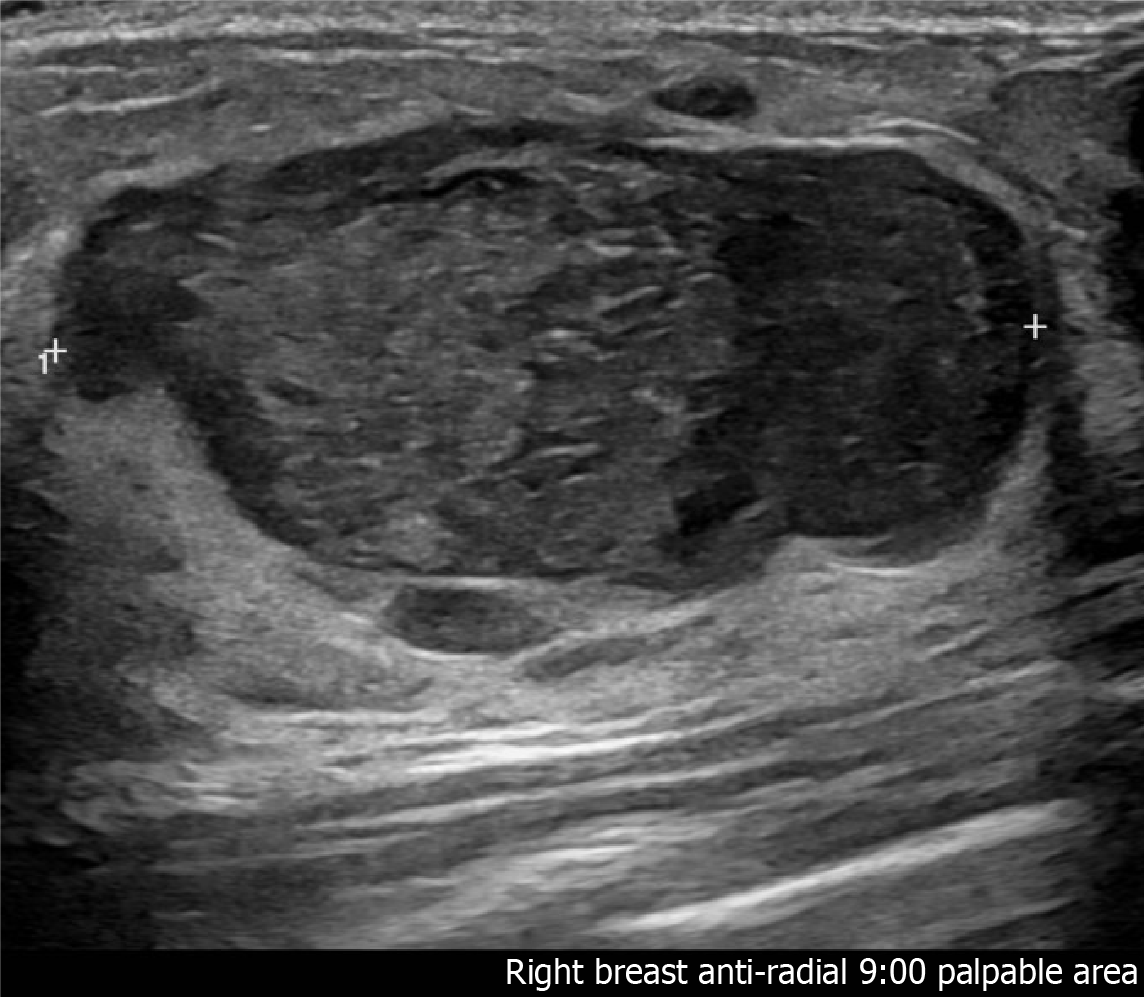
Figure 2 Initial breast ultrasound.
The radial (left) and anti-radial (right) scanning patterns showed a solid circumscribed oval mass in the right breast measuring 4.1 cm × 4.8 cm × 2.7 cm, suggestive of a fibroepithelial lesion, likely a benign fibroadenoma vs phyllodes tumor.
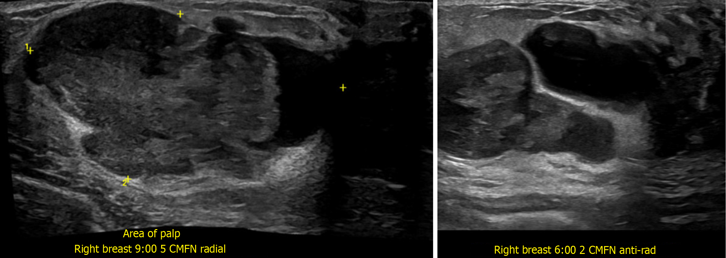
Figure 3 Repeat breast ultrasound two days after the initial presentation.
The radial (left) and anti-radial (right) scanning patterns showed enlargement of the mass to 5.3 cm × 2.9 cm × 5.5 cm and new cystic spaces with a peripheral cystic area measuring 2.4 cm. The rapid enlargement and development of cystic components suggest interval growth and necrosis, which raises the differential diagnosis of fibroepithelial lesion likely phyllodes tumor.
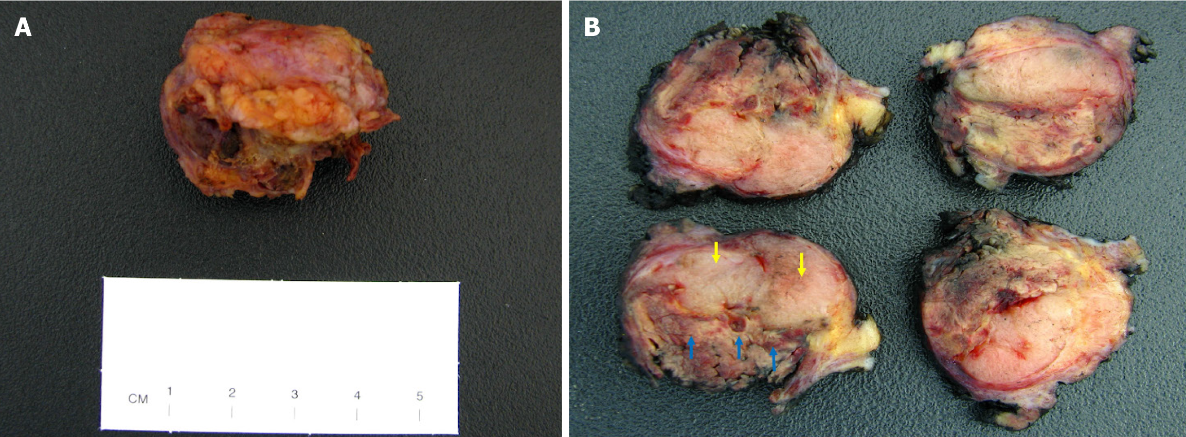
Figure 4 The gross appearance of the excision specimen.
A: A nodular fibrofatty tissue measuring 3.1 cm × 3 cm × 2.8 cm was received for pathologic evaluation; B: Serial sectioning of the specimen showed a well-circumscribed tan-white nodule measuring 23 cm in greatest dimension (yellow arrows). A portion of the nodule demonstrated areas of yellow discoloration with hemorrhage, consistent with infarction (blue arrows).
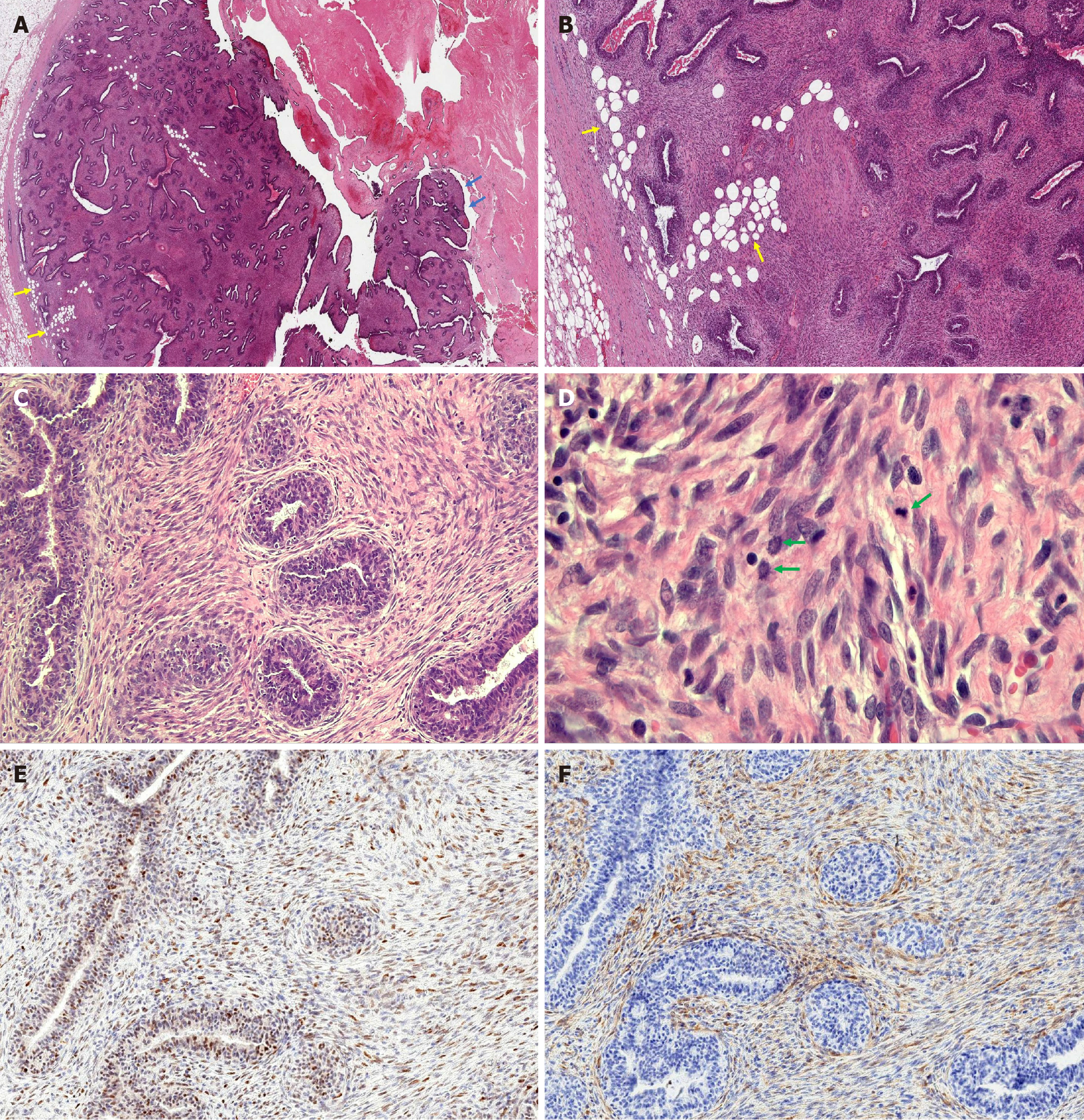
Figure 5 Microscopic features of the fibroepithelial lesion in the excision specimen.
A: Low magnification view showed a fibroepithelial neoplasm with focal papillary fronds growing into a cystic space (blue arrows). The tumor border was predominantly well-circumscribed with foci of stromal cells infiltrating the surrounding adipose tissue (yellow arrows), indicative of a microscopic permeative border. The glands displayed predominantly a pericanalicular growth pattern. Necrotic tissue consistent with infarction (top right) was also identified, corresponding to the gross tumor appearance. (H&E, 1 ×) B: Low magnification demonstrated increased stromal cellularity and permeative tumor border indicated by yellow arrows (H&E, 2 ×). C: Higher magnification showed increased stromal cellularity with mild periductal condensation (H&E, 10 ×). D: Plump stromal cells displayed mild to moderate cytologic atypia with three mitotic figures (green arrows) identified in one high-power field (H&E, 20 ×). E: p53 stain shows wild-type expression in the stromal cells (IHC, 10 ×). F: p16 stain shows normal patchy expression in the stromal cells (IHC, 10 ×).
- Citation: Suschana E, Sta Ines FM, Manrai P, Koelliker S, Gass JS, Tseng YA. Diagnostic and management challenges in a partially infarcted borderline phyllodes tumor in an adolescent female: A case report and review of literature. World J Clin Pediatr 2025; 14(3): 102741
- URL: https://www.wjgnet.com/2219-2808/full/v14/i3/102741.htm
- DOI: https://dx.doi.org/10.5409/wjcp.v14.i3.102741


