Copyright
©The Author(s) 2025.
World J Clin Pediatr. Sep 9, 2025; 14(3): 102741
Published online Sep 9, 2025. doi: 10.5409/wjcp.v14.i3.102741
Published online Sep 9, 2025. doi: 10.5409/wjcp.v14.i3.102741
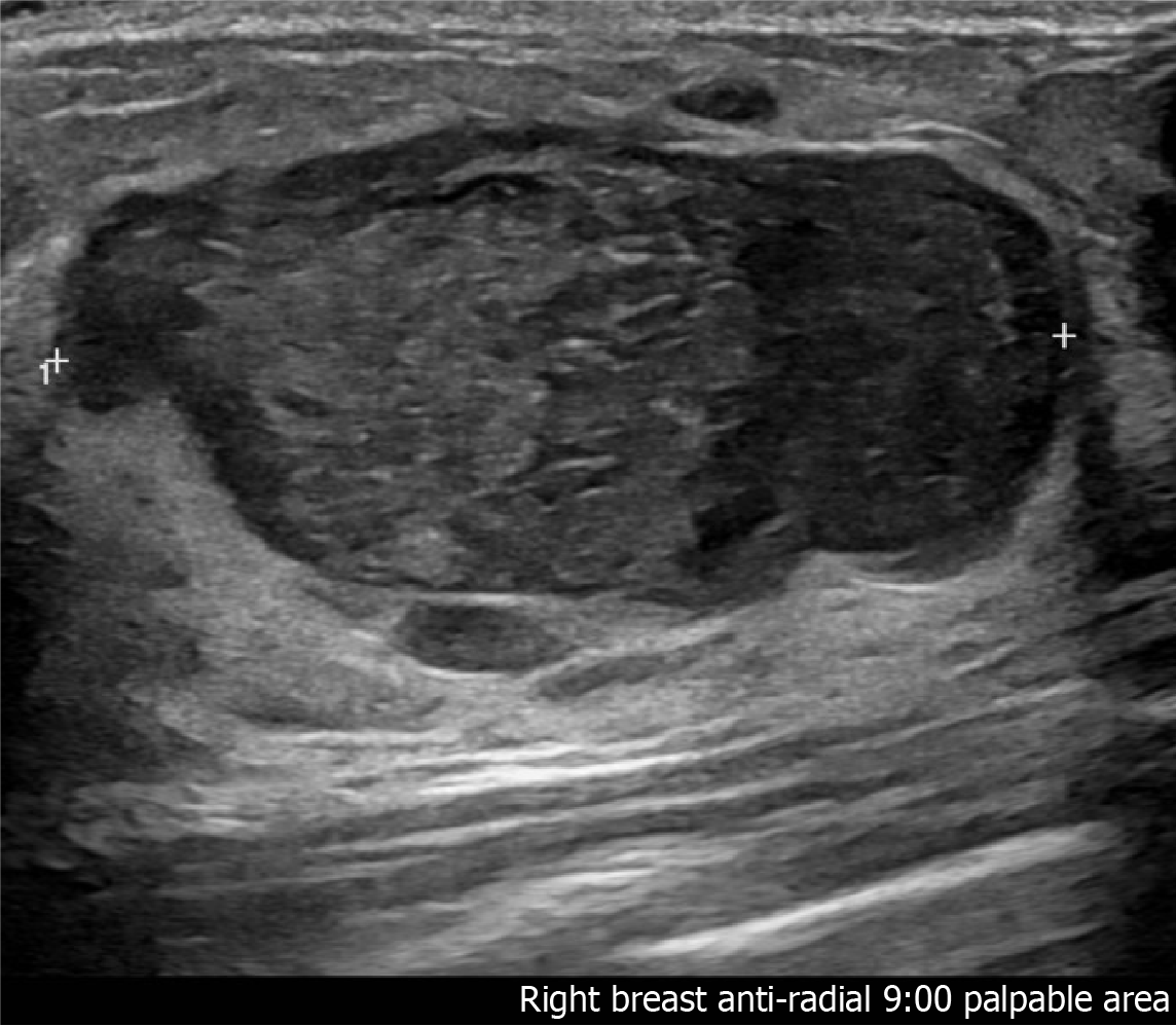
Figure 2 Initial breast ultrasound.
The radial (left) and anti-radial (right) scanning patterns showed a solid circumscribed oval mass in the right breast measuring 4.1 cm × 4.8 cm × 2.7 cm, suggestive of a fibroepithelial lesion, likely a benign fibroadenoma vs phyllodes tumor.
- Citation: Suschana E, Sta Ines FM, Manrai P, Koelliker S, Gass JS, Tseng YA. Diagnostic and management challenges in a partially infarcted borderline phyllodes tumor in an adolescent female: A case report and review of literature. World J Clin Pediatr 2025; 14(3): 102741
- URL: https://www.wjgnet.com/2219-2808/full/v14/i3/102741.htm
- DOI: https://dx.doi.org/10.5409/wjcp.v14.i3.102741


