Copyright
©The Author(s) 2025.
World J Clin Pediatr. Sep 9, 2025; 14(3): 102741
Published online Sep 9, 2025. doi: 10.5409/wjcp.v14.i3.102741
Published online Sep 9, 2025. doi: 10.5409/wjcp.v14.i3.102741
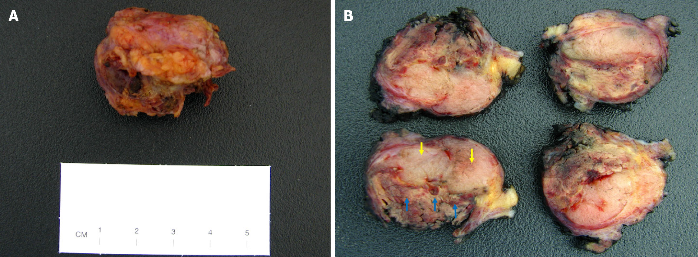
Figure 4 The gross appearance of the excision specimen.
A: A nodular fibrofatty tissue measuring 3.1 cm × 3 cm × 2.8 cm was received for pathologic evaluation; B: Serial sectioning of the specimen showed a well-circumscribed tan-white nodule measuring 23 cm in greatest dimension (yellow arrows). A portion of the nodule demonstrated areas of yellow discoloration with hemorrhage, consistent with infarction (blue arrows).
- Citation: Suschana E, Sta Ines FM, Manrai P, Koelliker S, Gass JS, Tseng YA. Diagnostic and management challenges in a partially infarcted borderline phyllodes tumor in an adolescent female: A case report and review of literature. World J Clin Pediatr 2025; 14(3): 102741
- URL: https://www.wjgnet.com/2219-2808/full/v14/i3/102741.htm
- DOI: https://dx.doi.org/10.5409/wjcp.v14.i3.102741


