Copyright
©The Author(s) 2025.
World J Clin Pediatr. Sep 9, 2025; 14(3): 102741
Published online Sep 9, 2025. doi: 10.5409/wjcp.v14.i3.102741
Published online Sep 9, 2025. doi: 10.5409/wjcp.v14.i3.102741
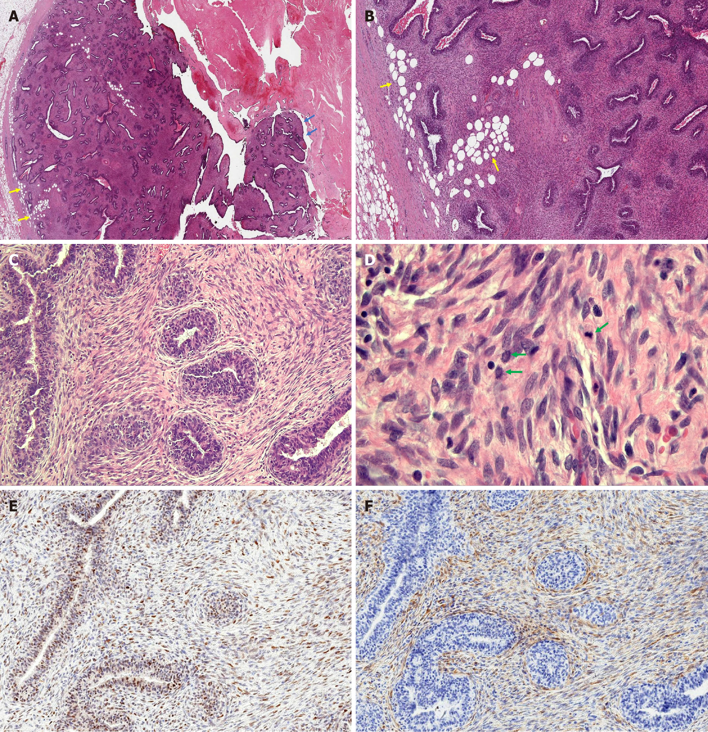
Figure 5 Microscopic features of the fibroepithelial lesion in the excision specimen.
A: Low magnification view showed a fibroepithelial neoplasm with focal papillary fronds growing into a cystic space (blue arrows). The tumor border was predominantly well-circumscribed with foci of stromal cells infiltrating the surrounding adipose tissue (yellow arrows), indicative of a microscopic permeative border. The glands displayed predominantly a pericanalicular growth pattern. Necrotic tissue consistent with infarction (top right) was also identified, corresponding to the gross tumor appearance. (H&E, 1 ×) B: Low magnification demonstrated increased stromal cellularity and permeative tumor border indicated by yellow arrows (H&E, 2 ×). C: Higher magnification showed increased stromal cellularity with mild periductal condensation (H&E, 10 ×). D: Plump stromal cells displayed mild to moderate cytologic atypia with three mitotic figures (green arrows) identified in one high-power field (H&E, 20 ×). E: p53 stain shows wild-type expression in the stromal cells (IHC, 10 ×). F: p16 stain shows normal patchy expression in the stromal cells (IHC, 10 ×).
- Citation: Suschana E, Sta Ines FM, Manrai P, Koelliker S, Gass JS, Tseng YA. Diagnostic and management challenges in a partially infarcted borderline phyllodes tumor in an adolescent female: A case report and review of literature. World J Clin Pediatr 2025; 14(3): 102741
- URL: https://www.wjgnet.com/2219-2808/full/v14/i3/102741.htm
- DOI: https://dx.doi.org/10.5409/wjcp.v14.i3.102741


