Minireviews Open Access
Copyright ©The Author(s) 2017. Published by Baishideng Publishing Group Inc. All rights reserved.
World J Orthop. Jan 18, 2017; 8(1): 12-20
Published online Jan 18, 2017. doi: 10.5312/wjo.v8.i1.12
Current management of talar osteochondral lesions
Arianna L Gianakos, Department of Orthopedic Surgery, 2nd Jersey City Medical Center, Jersey City, NJ 07302, United States
Arianna L Gianakos, Youichi Yasui, Charles P Hannon, John G Kennedy, Department of Foot and Ankle, Hospital for Special Surgery, New York, NY 10021, United States
Youichi Yasui, Department of Orthopaedic Surgery, 2nd Teikyo University School of Medicine, Tokyo 173-8606, Japan
Charles P Hannon, 2nd Department of Orthopaedic Surgery, Rush University Medical Center (C.P.H.), Chicago, IL 60612, United States
Author contributions: All authors equally contributed to this paper with conception and design of the study, literature review and analysis, drafting and critical revision and editing, and final approval of the final version.
Conflict-of-interest statement: Kennedy JG is a consultant for Arteriocyte, Inc.; Kennedy JG has received research support from the Ohnell Family Foundation, Mr. and Mrs. Michael J Levitt, and Arteriocyte Inc.; Kennedy JG is a board member for the European Society of Sports Traumatology, Knee Surgery, and Arthroscopy, International Society for Cartilage Repair of the Ankle, American Orthopaedic Foot and Ankle Society Awards and Scholarships Committee, International Cartilage Repair Society finance board.
Open-Access: This article is an open-access article which was selected by an in-house editor and fully peer-reviewed by external reviewers. It is distributed in accordance with the Creative Commons Attribution Non Commercial (CC BY-NC 4.0) license, which permits others to distribute, remix, adapt, build upon this work non-commercially, and license their derivative works on different terms, provided the original work is properly cited and the use is non-commercial. See: http://creativecommons.org/licenses/by-nc/4.0/
Correspondence to: John G Kennedy, MD, MCh, MMSc, FRCS (Orth), Department of Foot and Ankle, Hospital for Special Surgery, 523 East 72nd Street, Suite 507, New York, NY 10021, United States. kennedyj@hss.edu
Telephone: +1-646-7978880 Fax: +1-646-797896
Received: June 16, 2016
Peer-review started: June 17, 2016
First decision: July 27, 2016
Revised: September 12, 2016
Accepted: October 17, 2016
Article in press: October 18, 2016
Published online: January 18, 2017

Abstract

Osteochondral lesions of the talus (OLT) occur in up to 70% of acute ankle sprains and fractures. OLT have become increasingly recognized with the advancements in cartilage-sensitive diagnostic imaging modalities. Although OLT may be treated nonoperatively, a number of surgical techniques have been described for patients whom surgery is indicated. Traditionally, treatment of symptomatic OLT have included either reparative procedures, such as bone marrow stimulation (BMS), or replacement procedures, such as autologous osteochondral transplantation (AOT). Reparative procedures are generally indicated for OLT < 150 mm2 in area. Replacement strategies are used for large lesions or after failed primary repair procedures. Although short- and medium-term results have been reported, long-term studies on OLT treatment strategies are lacking. Biological augmentation including platelet-rich plasma and concentrated bone marrow aspirate is becoming increasingly popular for the treatment of OLT to enhance the biological environment during healing. In this review, we describe the most up-to-date clinical evidence of surgical outcomes, as well as both the mechanical and biological concerns associated with BMS and AOT. In addition, we will review the recent evidence for biological adjunct therapies that aim to improve outcomes and longevity of both BMS and AOT procedures.

Key Words: Osteochondral lesions of talus, Comprehensive review, Diagnosis, Bone marrow stimulation, Autologous autograft transfer, Biologics

Core tip: Osteochondral lesions of the talus are often missed after acute ankle sprains and fractures. Magnetic resonance imaging is most sensitive in diagnosing these injuries. Bone marrow stimulation (BMS) is effective for lesions < 150 mm2 in area, but replacement procedures such as autologous osteochondral transplantation or allografts may be required for larger lesions or if BMS fails. Long term studies should attempt to determine the most effective treatment strategy and the critical defect strategy beyond which BMS will not work.


INTRODUCTION

Osteochondral lesions of the talus (OLT) can occur in up to 70% of acute ankle sprains and fractures[1]. OLT have become increasingly recognized with the advancements in cartilage-sensitive diagnostic imaging modalities such as magnetic resonance imaging (MRI). These lesions typically involve a component of the articular surface and/or subchondral bone (SCB)[2]. Although trauma is the primary etiology, non-traumatic causes have been reported including congenital factors, ligamentous laxity, spontaneous necrosis, steroid treatment, embolic disease, and endocrine abnormalities[2,3].

A systematic review by Zengerink et al[4] demonstrated that up to 50% of patients failed to resolve their symptoms by conservative treatment. Traditionally, treatment of symptomatic OLT have included either reparative or replacement surgical procedures. Typically, the decision to repair or replace is based primarily on lesion size. Reparative procedures, including bone marrow stimulation (BMS), are generally indicated for OLT < 15 mm in a diameter or 150 mm2 in area[5]. Replacement strategies, such as osteochondral autologous transplantation (AOT), are used for large lesions or failed primary repair procedures[6]. Although previous clinical literature has demonstrated good to excellent short- and mid-term clinical outcomes, there has been an increase in the concerns regarding the methodological quality of previous clinical studies and deterioration of the ankle joint following surgical interventions.

In this review, we describe the most up-to-date clinical evidence of surgical outcomes, as well as increasing concerns associated with BMS and AOT. In addition, we will review the recent evidence for biological adjunct therapies that have been used to improve outcomes and longevity of both BMS and AOT.

CLINICAL PRESENTATION AND DIAGNOSIS

Most OLT are a sequelae of ankle injuries. Unfortunately, there are no specific physical examination findings that can accurately assess and diagnose OLT, and up to 50% of patients have missed OLT on plain radiographs[7]. It is therefore important to have a high level of suspicion of OLT in patients who have persistent ankle joint pain and a history of ankle injuries.

Patients with OLT frequently present with non-specific chronic ankle pain. Associated symptoms may also include generalized ankle swelling, stiffness, and weakness, which is often exacerbated by prolonged weight-bearing or high impact activities[2]. In the physical examination, a patient’s complaint of tenderness or pain may be poorly localized and may not correspond with the location of the OLT[8]. Examiners should perform both anterior drawer and standard inversion maneuvers to detect concomitant lateral ankle instability, and they should also assess hindfoot malalignment, joint flexibility, and joint laxity.

Anteroposterior, mortise, and lateral ankle weight-bearing radiographs are useful when assessing joint alignment and other coexisting abnormalities such as osteophytes and loose bodies. However, more advanced imaging is often recommended, since plain radiographs have been shown to miss up to 50% of OLT[9]. Computed tomography (CT) has excellent ability to detect OLT, accounting for 0.81 sensitivity and 0.99 specificity[7]. Although CT is useful in obtaining detail about bony injury including the condition of SCB, concomitant osteophytes, and loose bodies, it lacks the ability to assess the cartilage compartment of OLT. MRI is the recommended imaging diagnostic modality, with 0.96 sensitivity and 0.96 specificity[7]. MRI is advantageous in that it can show both osseous and soft tissue pathologies that are frequently associated in OLT. Although several scoring systems based on the MRI have been developed for grading of OLT[10-15], it is unclear whether any classification can direct clinical decision making. Research by Ferkel et al[11] showed little correlation between MRI grading and clinical outcomes. In a prospective study of 120 ankles, Choi et al[12] also found no correlation between any radiological grading and clinical outcome.

TREATMENT
Conservative treatment

Non-operative treatment strategies in asymptomatic patients can include rest and/or restriction of activities along with the use of a non-steroidal anti-inflammatory drug[4]. A systematic review by Zengerink et al[4] reported that 45% of patients reported successful outcomes when treated with conservative treatment consisting of weight-bearing as tolerated. The authors also demonstrated that 53% of patients who underwent cast immobilization for at least 3 wk up to 4 mo reported successful clinical outcomes. However, success was determined based on symptomatic complaint rather than on the physiological healing of the OLT. In addition, the long-term outcome of these treatment strategies has yet to be established. Recent clinical studies have revealed that OLT of the ankle joint have higher levels of intra-articular inflammatory cytokines than normal ankle joint which may lead to progressive deterioration of global, as well as focal lesions over time[16].

Operative treatment

There are two basic techniques for operative treatment for OLT: Reparative including BMS and replacement procedures including AOT. The decision to either proceed with BMS or AOT is primarily determined by lesion size. Traditionally, lesions of smaller sizes (< 15 mm in diameter or < 150 mm2 in area) are treated with BMS, while larger lesions are treated with AOT[6]. In addition, there has been recent evidence recommending AOT for patients who previously failed BMS[17].

BMS

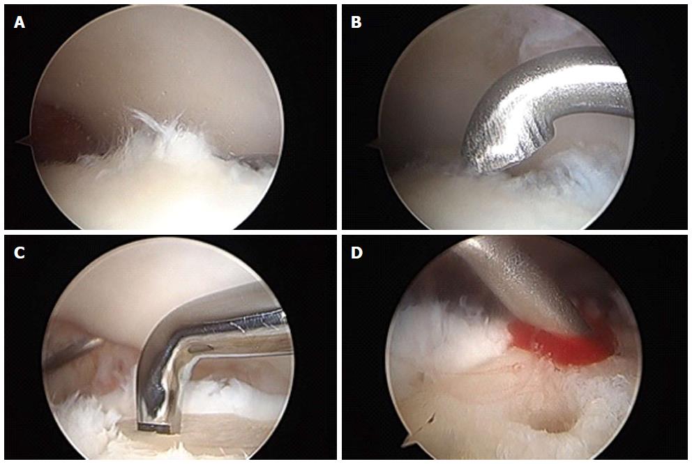
BMS is a reparative procedure that aims to stimulate the release of mesenchymal stem cells (MSCs) from the SCB marrow to infill fibrocartilage in the defect. In BMS, unstable cartilage, the calcified layer, and necrotic bone are debrided arthroscopically. A microfracture pick or small diameter drill is then used to penetrate the SCB plate (Figure 1).

Figure 1
Figure 1 Arthroscopic images of osteochondral lesions of the talus. A: Osteochondral lesion of the talus identified arthroscopically; B: Frayed or fibrillated cartilage is curretted out; C: Subchondral plate is violated with microfracture pick; D: After the subchondral bone plate is violated, bleeding occurs beginning the healing response.

While lesion size has been identified as the primary prognostic indicator affecting outcomes after BMS, several other prognostic factors have also been identified. Chuckpaiwong et al[5] reported that almost all patients in their series with OLT greater than 15 mm in diameter failed BMS (96.7%; 31/32) while the other patients with lesions less than 15 mm in diameter had 100% success. Choi et al[12] demonstrated a risk of failure with lesions greater than 150 mm2 on MRI. Another important prognostic factor is containment (shoulder vs non-shoulder type) of OLT. Choi et al[18] demonstrated that patients with shoulder-type OLT were more likely to have a worse clinical outcome than non-shoulder lesions. Because of the nature of BMS, subchondral bone cyst may affect the outcomes. To address this, Lee et al[19] performed a randomized control study and found that there were no significant differences in clinical outcomes between patients in the subchondral cyst group and those patients treated with no subchondral cyst component. However, the longevity of these outcomes is of concern due to the lack of mechanical and biological function of SCB required for robust cartilage repair[20].

Several clinical studies have demonstrated that nearly 85% of patients undergoing BMS report good to excellent clinical short- and mid-term outcomes[4,21]. van Bergen et al[22] evaluated long term clinical outcomes in 50 patients with at a mean follow-up of 141 mo and reported a mean American Orthopaedic Foot and Ankle Society (AOFAS) score of 88 out of 100 possible points. Polat et al[23] demonstrated that out of 82 patients treated with BMS, 42.6% of patients had no symptoms and 23.1% of patients had pain after walking more than 2 h or after competitive sports activities at a mean follow-up of 121.3 mo.

Despite successful outcomes following BMS for OLT, there have been numerous studies demonstrating cause for concerns including the quality of the studies reporting positive outcomes, mechanical concerns regarding the fibrocartilage repair tissue, and long-term deteriorating clinical outcomes[21]. A systematic review by Hannon et al[24] found gross inconsistencies and an underreporting of data in the included 24 clinical studies that report clinical outcomes after BMS for OLT. The authors found that only 46% of clinical studies reported the lesion size and only 25% performed postoperative radiological evaluation. Therefore, the authors concluded that there is not enough data in the current literature to accurately assess the outcome of BMS[24].

Deterioration of reparative fibrocartilage quality has been reported in up to 35% of patients within the first five years of BMS, and only 30% of patients who received BMS have integration of the repair tissue with the surrounding native cartilage at second look arthroscopy 12 mo postoperatively[11,14]. Becher et al[25] also demonstrated that although tissue regenerated at the site of microfracture, it was neither intact not homogeneous. In a series of 120 ankles, Choi et al[12] has shown deterioration of clinical success rate over time following BMS.

There are numerous factors that may play a role in affecting the durability of the repair tissue following BMS. There is an increased awareness that impairment of SCB following BMS may be a cause of deterioration. Anatomically, the SCB is located under the articular cartilage offering biomechanical and biological support for overlying articular cartilage[26,27]. During BMS, there is gross destruction of cross-talk between the SCB plate and the articular cartilage. This destruction is a result of the surgical trauma and compaction of the SCB plate that occurs with penetration of either a microfracture pic or drilling[27]. In the sheep osteochondral lesion model, Orth et al[28] revealed that the SCB plate was not restored at 6 mo after BMS. This finding was supported in the human ankle by Reilingh et al[29] which revealed that the SCB were not filled completely in 78.6% (44 of 58) OLT at 1 year after BMS. This inevitable trauma to the SCB may be limited by using a small diameter microfracture pic rather than drilling or using larger diameter conventional microfracture pics[27].

Mechanical and biological insufficiency may be part of the reasons for deterioration of fibrocartilage. Marrow stimulating techniques attempt to fill talar lesions with precursor cells and cytokines, resulting in a fibrin clot that will ultimately lead to fibrocartilaginous type-1 collagen formation[10,24]. This cartilage consists of collagen that has different biomechanical properties than the native hyaline cartilage containing type-II collagen. It has been demonstrated that fibrocartilage has inferior stiffness, resilience, and wear properties and therefore is at risk of degeneration[30,31].

AOT

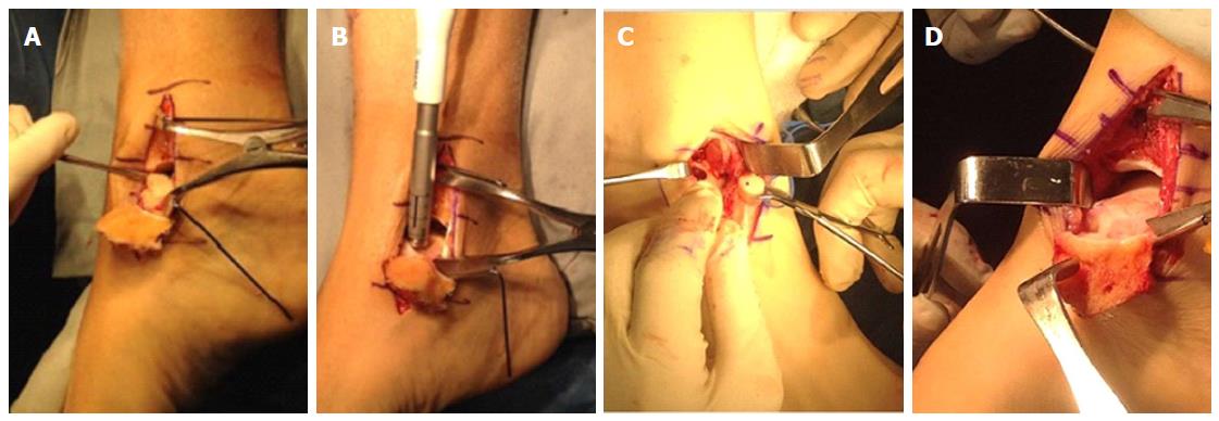
AOT replaces cartilage by transplanting a cylindrical osteochondral graft from a non weightbearing portion of the knee into a defect site on the talus (Figure 2). AOT is indicated in patients with lesion sizes greater than 15 mm in diameter or 150 mm2, or in cases of failed previous BMS[4,6]. Kim et al[32] reported prognostic factors affecting outcomes of AOT and found that patient age, sex, body mass index, duration of symptoms, location of OLT, and the existence of a subchondral cyst did not significantly influence clinical outcomes of AOT. By Haleem et al[33] reported that the size of the OLT is also not a significant predictor of outcomes and multiple grafts may be used without adversely affecting the outcome.

Figure 2
Figure 2 Autologous osteochondral transplantation procedure. A: Medial exposure of the talus; B: Preparation of the defect site; C: Insertion of cylindrical osteochondral plug into the prepared osteochondral lesions of the talus defect site; D: Exposure of the medial talus via the chevron-type medial malleolar osteotomy.

Several studies have reported good clinical outcomes following AOT at both short- and mid-term follow-up. A case series on 85 patients who underwent AOT found improved Foot and Ankle Outcome Score (FAOS) at 47.2 mo follow-up and improved Magnetic Resonance Observation of Cartilage Repair Tissue (MOCART) scores post-operatively at 24.8 mo follow-up[34]. One study by Haleem et al[33] compared clinical and radiological MRI outcomes of OLT treated by single-plug vs double-plug AOT at 5-year follow-up. They found treatment with double-plug AOT did not show inferior clinical or radiological outcomes when compared to single-plug AOT in the intermediate term. Good outcomes are not limited to the general population only, and excellent outcomes have been reported in the athletic population at midterm follow-up. Fraser et al[35] reported improved AOFAS scores and found at final follow up of 24 mo, 90% of professional athletes and 87% of recreational athletes were able to return to pre-injury activity levels. Despite its apparent success and favorable short- and medium-term outcome profile, there has been no study to our knowledge that has described long-term (10+ years) outcomes after AOT.

AOT outcome studies however should be evaluated carefully. Hannon et al[24] showed that outcomes and clinical variables were reported in less than 73% and 67% of studies respectively. Therefore, the data between studies reported have been incongruent and limit cross sectional comparison

AOT has good clinical outcomes, but there are some mechanical concerns with the procedure such as formation of post-operative cysts, morbidity associated with accessing the ankle joint through osteotomies, and pressures on the graft due to malalignment. It has been suggested that biomechanical success may be limited by the alignment of the graft. Fansa et al[36] demonstrated increased contact pressure on the graft surface by 7-fold with a 1.0 mm of graft protrusion above the level of the native cartilage. Other mechanical considerations have also been an area of concern with AOT. The use of a medial malleolar osteotomy has raised concerns for increasing the risk of mal/non-union. However, current evidence suggests adequate osteotomy, both medially and laterally, as well as cartilaginous healing in the short- to mid-term follow-up. Lamb et al[37] demonstrated that a Chevron-type medial malleolar osteotomy had overall improved healing and fixation, with evidence of fibrocartilaginous tissue present at the superficial osteotomy interface. In addition, at a mean follow-up of 64 mo, a retrospective case series by Gianakos et al[38] demonstrated that an anterolateral tibial osteotomy resulted in T2 mapping relaxation times similar to both superficial and deep interfaces of the native cartilage and had overall improved FAOS and MOCART scores and. However, it is known that ankle fractures may cause activation of intra-articular inflammatory cytokines, which may lead to progressive deterioration of OLT over time, and this may theoretically occur with malleolar osteotomy[16]. There have been reports demonstrating the potential of poor integration of the AOT surface with the native tissue, cyst formation around the graft site, and deterioration of the graft cartilage as potential consequences following AOT procedure. However, a case series by Savage-Elliott et al[39] demonstrated that although increasing age was related to increased cyst prevalence, the clinical impact of cyst formation was not found to be significant at a mean short-term follow up of 15 mo after surgery.

Lastly, concerns over donor site morbidity have gained increasing attention. Valderrabano et al[40] reported on the outcomes of 12 patients undergoing AOT, of whom 50% experienced donor site morbidity with all patients showing MRI signs of cartilage change, joint space narrowing, or cystic changes in untreated donor sites. These results have been challenged by similar reports. Yoon et al[17] found in 22 patients a 9% early donor site morbidity with 100% resolution at 48 mo follow-up. Fraser et al[41] performed a retrospective analysis on 39 patients who underwent AOT and reported that at 24 mo follow-up, donor site morbidity was present in only 5% of patients and that Lysholm scores were at 99.4 for the entire cohort. Therefore, OLT treated with AOT can have a low incidence of donor site morbidity with good functional outcomes.

Although the overall success of AOT for OLT may be limited by a combination of factors, evidence in the literature suggests that AOT is effective short- and mid-term follow-up, particularly for large lesions that may not be managed by other forms of treatment.

Osteochondral allograft transplantation

Osteochondral allograft transplantation is a technique that has been employed for the treatment of OLT and involves replacing defects in bone and articular cartilage with cadaveric donor specimens[42]. Some surgeons prefer this procedure over AOT because it avoids donor site morbidity[24]. Although frozen grafts may be used, the decline in the viability of chondrocytes within the graft tissue has led to an increase in the use of fresh allografts.

Reported success rates are highly variable within the literature. El-Rashidy et al[43] performed one of the largest studies published on patients who received small cylindrical allografts and reported positive outcomes in 28 of 38 patients at a mean follow-up of 37.7 mo. Raikin[44] evaluated patients who received bulk allografts and demonstrated improved AOFAS scores in 15 patients at a mean follow-up of 44 mo. Lastly, Haene et al[45] reported in a case series that only ten of 17 cases who underwent allograft transplantation had good or excellent results at an average follow-up of 4.1 years. Although clinical evidence suggests osteochondral allograft transplantation to be effective in the treatment of larger OLT, this evidence is limited as it consists primarily of case series with reported variable success rates.

Autologous chondrocyte implantation

Autologous chondrocyte implantation (ACI) is a cell-based, two-stage procedure that can be used as an alternative to osteochondral grafting techniques. This technique involves harvesting healthy articular cartilage for chondrocyte cultures, which are grown for approximately 30 d[46]. These cultures are implanted into the defect site. The aim of ACI is to promote the development of hyaline-like repair tissue. ACI is typically indicated for full-thickness cartilage defects with an intact SCB plate with stable edges of the surrounding cartilage[47].

A systematic review by Harris et al[48] analyzed 82 studies (5276 subjects; 6080 defects) and reported a low failure rate of 1.5%-7.7% following ACI in the knee. Similar outcomes have been shown in the ankle. A meta-analysis by Niemeyer et al[49] reported a clinical success rate of 89.9% in 213 patients following ACI. Gobbi et al[50] reported no difference in AOFAS scores following chondroplasty, microfracture, and AOT. Disadvantages of ACI include the cost of culturing hyaline cells, the need for two surgical procedures, hypertrophy of the graft and the durability of the graft[2].

Although many studies have published promising results, the available evidence to date is of poor quality due to the level of evidence, low patient number, and use of variable outcome parameters[47]. Therefore, randomized clinical trials are necessary to determine the superiority of ACI over other more established techniques.

Matrix-induced autologous chondrocyte implantation

Matrix-induced autologous chondrocyte implantation (MACI) is a second generation of ACI whereby cells are embedded into a bioabsorbable matrix[24]. This membrane is placed over the talar cartilage defect. This procedure avoids periosteal graft harvesting and allows for a more even cell distribution[51]. In addition, a fibrin sealant can be utilized to secure the defect, reducing the need for suture fixation.

Evidence in the literature has demonstrated arthroscopic MACI as a safe alternative for the treatment of OLT with good overall clinical and radiologic results. Aurich et al[52] reported in a case series of 19 patients, significant improvement in AOFAS clinical scores following MACI at a mean follow-up of 24 mo. Giannini et al[53] also reported positive clinical and histologic outcome scores at 36 mo post-operatively.

Evidence has demonstrated MACI to be a promising new treatment method for large OLT. Future research should attempt to compare radiological, clinical, and histological MACI to conventional treatment.

BIOLOGIC AUGUMENTATION FOR CARTILAGE REPAIR
Platelet-rich plasma

Platelet-rich plasma (PRP) is an autologous blood product that contains at least twice the concentration of platelets compared to baseline values, or > 1.1 × 10⁶ platelets/μL[54]. Platelets contain numerous growth factors and cytokines which have been shown to induce human-MSC proliferation and promote tissue healing[55].

There has been evidence in the literature that demonstrates positive effects of PRP on cartilage repair. Smyth et al[56] showed in a systematic review that 18 of 21 (85.7%) basic science papers reported positive effects of PRP on cartilage repair. Additionally, Smyth et al[57] found in a rabbit model, that application of PRP at time of AOT improved the integration of the osteochondral graft at the cartilage interface and decreased graft degeneration. In clinical studies, Guney et al[58] performed a randomized control trial in 19 OLT patients and reported that BMS with PRP had better functional outcomes when compared with BMS alone. Görmeli et al[59] compared the effect of PRP and HA following BMS for OLT and found at 15.3-mo follow-up, clinical improvement after PRP with HA when compared to HA or saline injection alone.

Despite successful reported outcome following PRP adjuvants, the effect of PRP on OLT is still controversial because of several concerns. Currently there has been no proposed standard method for PRP harvesting. There are a variety of commercially-available centrifugation systems with various timing protocols and activation methods[60]. In addition, plasma contains differing concentrations of platelets, cells, growth factors, and cytokine, which are variable even within a single individual[60]. Several studies have evaluated the anti-inflammatory effects of different leukocyte concentrated PRP on cartilage repair[61,62]. However, to our knowledge, there has be no study that has investigated the effect of leukocyte concentration in PRP in the treatment of ankle OLT. In conclusion, the published literature suggests that utilizing PRP in the operative treatment for OLT can improve clinical and functional outcomes. The evidence for PRP is promising; however, well-designed clinical trials are necessary to determine its efficacy in the clinical setting.

Concentrated bone marrow aspirate

Concentrated Bone Marrow Aspirate (cBMA) is a blood product produced by centrifuging bone marrow typically aspirated from the iliac crest[63]. cBMA contains a variety of bioactive cytokines, as well as MSCs, which have the ability to undergo chondrocyte differentiation. In addition, most recent studies have shown that cBMA includes an abundant concentration of interleukin-1 receptor antagonist proteins (IL-1Ra), which are the primary anti-inflammatory cytokines[63].

A few studies have demonstrated the ability of cBMA to promote the chondrogenic cascade which can be beneficial in the treatment of osteochondral lesions. Improved cartilage healing has been demonstrated in the equine model, with improvements histologically and radiographically in groups receiving cBMA at the time of BMS[64]. In addition, similar results were reported in a goat model when using BMS combination with cBMA and HA[65]. Clinically, Hannon et al[66] reported that mean FAOS improved significantly pre- to post-operatively at 48.3 mo in groups receiving cBMA with BMS. They also demonstrated that groups with cBMA had improved integration of the repair tissue with MRI demonstrating less fissuring and fibrillation. Kennedy et al[6] demonstrated improved restoration of radius curvature and color stratification similar to that of native cartilage on MRI using T2 mapping in patients treated with cBMA and AOT. Overall, current evidence suggests that cBMA can improve cartilage repair in OLT, but future clinical research and clinical trials are necessary for better comparison of outcomes with other biological adjuncts.

CONCLUSION

OLT present a challenge and optimal treatment remains controversial. Although future randomized clinical trials are needed to establish evidence of the most effective treatment, both reparative and replacement procedures remain feasible options. The literature supports treatment with BMS for lesions of smaller sizes, whereas treatment with AOT may be utilized for larger or cystic lesions. Cell-based techniques and allograft transplantation may be utilized in failed primary procedures. Although biologic augmentation offers promising results, well-designed clinical trials are necessary to determine efficacy in the clinical setting.

Footnotes

Manuscript source: Invited manuscript

Specialty type: Orthopedics

Country of origin: United States

Peer-review report classification

Grade A (Excellent): 0

Grade B (Very good): B, B, B

Grade C (Good): C

Grade D (Fair): 0

Grade E (Poor): 0

P- Reviewer: Angoules A, Fenichel I, Guerado E, Ohishi T S- Editor: Gong XM L- Editor: A E- Editor: Wu HL

References
1.  Hintermann B, Regazzoni P, Lampert C, Stutz G, Gächter A. Arthroscopic findings in acute fractures of the ankle. J Bone Joint Surg Br. 2000;82:345-351.  [PubMed]  [DOI]  [Cited in This Article: ]  [Cited by in Crossref: 126]  [Cited by in F6Publishing: 151]  [Article Influence: 6.3]  [Reference Citation Analysis (0)]
2.  O’Loughlin PF, Heyworth BE, Kennedy JG. Current concepts in the diagnosis and treatment of osteochondral lesions of the ankle. Am J Sports Med. 2010;38:392-404.  [PubMed]  [DOI]  [Cited in This Article: ]  [Cited by in Crossref: 223]  [Cited by in F6Publishing: 194]  [Article Influence: 13.9]  [Reference Citation Analysis (0)]
3.  Gardiner TB. Osteochondritis dissecans in three members of one family. J Bone Joint Surg Br. 1955;37-B:139-141.  [PubMed]  [DOI]  [Cited in This Article: ]
4.  Zengerink M, Szerb I, Hangody L, Dopirak RM, Ferkel RD, van Dijk CN. Current concepts: treatment of osteochondral ankle defects. Foot Ankle Clin. 2006;11:331-359, vi.  [PubMed]  [DOI]  [Cited in This Article: ]  [Cited by in Crossref: 127]  [Cited by in F6Publishing: 91]  [Article Influence: 5.1]  [Reference Citation Analysis (0)]
5.  Chuckpaiwong B, Berkson EM, Theodore GH. Microfracture for osteochondral lesions of the ankle: outcome analysis and outcome predictors of 105 cases. Arthroscopy. 2008;24:106-112.  [PubMed]  [DOI]  [Cited in This Article: ]  [Cited by in Crossref: 342]  [Cited by in F6Publishing: 312]  [Article Influence: 19.5]  [Reference Citation Analysis (0)]
6.  Kennedy JG, Murawski CD. The Treatment of Osteochondral Lesions of the Talus with Autologous Osteochondral Transplantation and Bone Marrow Aspirate Concentrate: Surgical Technique. Cartilage. 2011;2:327-336.  [PubMed]  [DOI]  [Cited in This Article: ]  [Cited by in Crossref: 114]  [Cited by in F6Publishing: 118]  [Article Influence: 9.1]  [Reference Citation Analysis (0)]
7.  Verhagen RA, Maas M, Dijkgraaf MG, Tol JL, Krips R, van Dijk CN. Prospective study on diagnostic strategies in osteochondral lesions of the talus. Is MRI superior to helical CT? J Bone Joint Surg Br. 2005;87:41-46.  [PubMed]  [DOI]  [Cited in This Article: ]
8.  Stone JW Debridement with stimulation of the base. The Foot and Ankle: AANA Advanced Arthroscopic Surgical Techniques 2016; 9-17.  [PubMed]  [DOI]  [Cited in This Article: ]
9.  Loomer R, Fisher C, Lloyd-Smith R, Sisler J, Cooney T. Osteochondral lesions of the talus. Am J Sports Med. 1993;21:13-19.  [PubMed]  [DOI]  [Cited in This Article: ]  [Cited by in Crossref: 265]  [Cited by in F6Publishing: 275]  [Article Influence: 8.9]  [Reference Citation Analysis (0)]
10.  McGahan PJ, Pinney SJ. Current concept review: osteochondral lesions of the talus. Foot Ankle Int. 2010;31:90-101.  [PubMed]  [DOI]  [Cited in This Article: ]  [Cited by in Crossref: 101]  [Cited by in F6Publishing: 99]  [Article Influence: 7.1]  [Reference Citation Analysis (0)]
11.  Ferkel RD, Zanotti RM, Komenda GA, Sgaglione NA, Cheng MS, Applegate GR, Dopirak RM. Arthroscopic treatment of chronic osteochondral lesions of the talus: long-term results. Am J Sports Med. 2008;36:1750-1762.  [PubMed]  [DOI]  [Cited in This Article: ]  [Cited by in Crossref: 325]  [Cited by in F6Publishing: 333]  [Article Influence: 20.8]  [Reference Citation Analysis (0)]
12.  Choi WJ, Park KK, Kim BS, Lee JW. Osteochondral lesion of the talus: is there a critical defect size for poor outcome? Am J Sports Med. 2009;37:1974-1980.  [PubMed]  [DOI]  [Cited in This Article: ]  [Cited by in Crossref: 313]  [Cited by in F6Publishing: 323]  [Article Influence: 21.5]  [Reference Citation Analysis (0)]
13.  Mintz DN, Tashjian GS, Connell DA, Deland JT, O’Malley M, Potter HG. Osteochondral lesions of the talus: a new magnetic resonance grading system with arthroscopic correlation. Arthroscopy. 2003;19:353-359.  [PubMed]  [DOI]  [Cited in This Article: ]  [Cited by in Crossref: 166]  [Cited by in F6Publishing: 169]  [Article Influence: 8.0]  [Reference Citation Analysis (0)]
14.  Lee KB, Bai LB, Park JG, Yoon TR. A comparison of arthroscopic and MRI findings in staging of osteochondral lesions of the talus. Knee Surg Sports Traumatol Arthrosc. 2008;16:1047-1051.  [PubMed]  [DOI]  [Cited in This Article: ]  [Cited by in Crossref: 32]  [Cited by in F6Publishing: 33]  [Article Influence: 2.1]  [Reference Citation Analysis (0)]
15.  Hepple S, Winson IG, Glew D. Osteochondral lesions of the talus: a revised classification. Foot Ankle Int. 1999;20:789-793.  [PubMed]  [DOI]  [Cited in This Article: ]  [Cited by in Crossref: 260]  [Cited by in F6Publishing: 197]  [Article Influence: 7.9]  [Reference Citation Analysis (0)]
16.  Henkelmann R, Schmal H, Pilz IH, Salzmann GM, Dovi-Akue D, Südkamp NP. Prospective clinical trial of patients who underwent ankle arthroscopy with articular diseases to match clinical and radiological scores with intra-articular cytokines. Int Orthop. 2015;39:1631-1637.  [PubMed]  [DOI]  [Cited in This Article: ]  [Cited by in Crossref: 6]  [Cited by in F6Publishing: 6]  [Article Influence: 0.7]  [Reference Citation Analysis (0)]
17.  Yoon HS, Park YJ, Lee M, Choi WJ, Lee JW. Osteochondral Autologous Transplantation Is Superior to Repeat Arthroscopy for the Treatment of Osteochondral Lesions of the Talus After Failed Primary Arthroscopic Treatment. Am J Sports Med. 2014;42:1896-1903.  [PubMed]  [DOI]  [Cited in This Article: ]  [Cited by in Crossref: 55]  [Cited by in F6Publishing: 56]  [Article Influence: 5.6]  [Reference Citation Analysis (0)]
18.  Choi WJ, Choi GW, Kim JS, Lee JW. Prognostic significance of the containment and location of osteochondral lesions of the talus: independent adverse outcomes associated with uncontained lesions of the talar shoulder. Am J Sports Med. 2013;41:126-133.  [PubMed]  [DOI]  [Cited in This Article: ]  [Cited by in Crossref: 71]  [Cited by in F6Publishing: 72]  [Article Influence: 6.5]  [Reference Citation Analysis (0)]
19.  Lee KB, Park HW, Cho HJ, Seon JK. Comparison of Arthroscopic Microfracture for Osteochondral Lesions of the Talus With and Without Subchondral Cyst. Am J Sports Med. 2015;43:1951-1956.  [PubMed]  [DOI]  [Cited in This Article: ]  [Cited by in Crossref: 38]  [Cited by in F6Publishing: 38]  [Article Influence: 4.2]  [Reference Citation Analysis (0)]
20.  Goldring SR. Alterations in periarticular bone and cross talk between subchondral bone and articular cartilage in osteoarthritis. Ther Adv Musculoskelet Dis. 2012;4:249-258.  [PubMed]  [DOI]  [Cited in This Article: ]  [Cited by in Crossref: 117]  [Cited by in F6Publishing: 122]  [Article Influence: 10.2]  [Reference Citation Analysis (0)]
21.  Murawski CD, Kennedy JG. Operative treatment of osteochondral lesions of the talus. J Bone Joint Surg Am. 2013;95:1045-1054.  [PubMed]  [DOI]  [Cited in This Article: ]  [Cited by in Crossref: 186]  [Cited by in F6Publishing: 160]  [Article Influence: 14.5]  [Reference Citation Analysis (0)]
22.  van Bergen CJ, Kox LS, Maas M, Sierevelt IN, Kerkhoffs GM, van Dijk CN. Arthroscopic treatment of osteochondral defects of the talus: outcomes at eight to twenty years of follow-up. J Bone Joint Surg Am. 2013;95:519-525.  [PubMed]  [DOI]  [Cited in This Article: ]  [Cited by in Crossref: 138]  [Cited by in F6Publishing: 118]  [Article Influence: 10.7]  [Reference Citation Analysis (0)]
23.  Polat G, Erşen A, Erdil ME, Kızılkurt T, Kılıçoğlu Ö, Aşık M. Long-term results of microfracture in the treatment of talus osteochondral lesions. Knee Surg Sports Traumatol Arthrosc. 2016;24:1299-1303.  [PubMed]  [DOI]  [Cited in This Article: ]  [Cited by in Crossref: 87]  [Cited by in F6Publishing: 88]  [Article Influence: 11.0]  [Reference Citation Analysis (0)]
24.  Hannon CP, Smyth NA, Murawski CD, Savage-Elliott I, Deyer TW, Calder JD, Kennedy JG. Osteochondral lesions of the talus: aspects of current management. Bone Joint J. 2014;96-B:164-171.  [PubMed]  [DOI]  [Cited in This Article: ]  [Cited by in Crossref: 113]  [Cited by in F6Publishing: 97]  [Article Influence: 9.7]  [Reference Citation Analysis (0)]
25.  Becher C, Driessen A, Hess T, Longo UG, Maffulli N, Thermann H. Microfracture for chondral defects of the talus: maintenance of early results at midterm follow-up. Knee Surg Sports Traumatol Arthrosc. 2010;18:656-663.  [PubMed]  [DOI]  [Cited in This Article: ]  [Cited by in Crossref: 120]  [Cited by in F6Publishing: 125]  [Article Influence: 8.9]  [Reference Citation Analysis (0)]
26.  Sharma AR, Jagga S, Lee SS, Nam JS. Interplay between cartilage and subchondral bone contributing to pathogenesis of osteoarthritis. Int J Mol Sci. 2013;14:19805-19830.  [PubMed]  [DOI]  [Cited in This Article: ]  [Cited by in Crossref: 177]  [Cited by in F6Publishing: 185]  [Article Influence: 16.8]  [Reference Citation Analysis (0)]
27.  Gianakos AL, Yasui Y, Fraser EJ, Ross KA, Prado MP, Fortier LA, Kennedy JG. The Effect of Different Bone Marrow Stimulation Techniques on Human Talar Subchondral Bone: A Micro-Computed Tomography Evaluation. Arthroscopy. 2016;32:2110-2117.  [PubMed]  [DOI]  [Cited in This Article: ]  [Cited by in Crossref: 22]  [Cited by in F6Publishing: 22]  [Article Influence: 2.8]  [Reference Citation Analysis (0)]
28.  Orth P, Meyer HL, Goebel L, Eldracher M, Ong MF, Cucchiarini M, Madry H. Improved repair of chondral and osteochondral defects in the ovine trochlea compared with the medial condyle. J Orthop Res. 2013;31:1772-1779.  [PubMed]  [DOI]  [Cited in This Article: ]  [Cited by in Crossref: 45]  [Cited by in F6Publishing: 46]  [Article Influence: 4.2]  [Reference Citation Analysis (0)]
29.  Reilingh ML, van Bergen CJ, Blankevoort L, Gerards RM, van Eekeren IC, Kerkhoffs GM, van Dijk CN. Computed tomography analysis of osteochondral defects of the talus after arthroscopic debridement and microfracture. Knee Surg Sports Traumatol Arthrosc. 2016;24:1286-1292.  [PubMed]  [DOI]  [Cited in This Article: ]  [Cited by in Crossref: 30]  [Cited by in F6Publishing: 29]  [Article Influence: 3.6]  [Reference Citation Analysis (0)]
30.  Furukawa T, Eyre DR, Koide S, Glimcher MJ. Biochemical studies on repair cartilage resurfacing experimental defects in the rabbit knee. J Bone Joint Surg Am. 1980;62:79-89.  [PubMed]  [DOI]  [Cited in This Article: ]
31.  Nehrer S, Spector M, Minas T. Histologic analysis of tissue after failed cartilage repair procedures. Clin Orthop Relat Res. 1999;149-162.  [PubMed]  [DOI]  [Cited in This Article: ]  [Cited by in Crossref: 295]  [Cited by in F6Publishing: 297]  [Article Influence: 11.9]  [Reference Citation Analysis (0)]
32.  Kim YS, Park EH, Kim YC, Koh YG, Lee JW. Factors associated with the clinical outcomes of the osteochondral autograft transfer system in osteochondral lesions of the talus: second-look arthroscopic evaluation. Am J Sports Med. 2012;40:2709-2719.  [PubMed]  [DOI]  [Cited in This Article: ]  [Cited by in Crossref: 65]  [Cited by in F6Publishing: 66]  [Article Influence: 5.5]  [Reference Citation Analysis (0)]
33.  Haleem AM, Ross KA, Smyth NA, Duke GL, Deyer TW, Do HT, Kennedy JG. Double-Plug Autologous Osteochondral Transplantation Shows Equal Functional Outcomes Compared With Single-Plug Procedures in Lesions of the Talar Dome: A Minimum 5-Year Clinical Follow-up. Am J Sports Med. 2014;42:1888-1895.  [PubMed]  [DOI]  [Cited in This Article: ]  [Cited by in Crossref: 35]  [Cited by in F6Publishing: 35]  [Article Influence: 3.5]  [Reference Citation Analysis (0)]
34.  Flynn S, Ross KA, Hannon CP, Yasui Y, Newman H, Murawski CD, Deyer TW, Do HT, Kennedy JG. Autologous Osteochondral Transplantation for Osteochondral Lesions of the Talus. Foot Ankle Int. 2016;37:363-372.  [PubMed]  [DOI]  [Cited in This Article: ]  [Cited by in Crossref: 39]  [Cited by in F6Publishing: 40]  [Article Influence: 5.0]  [Reference Citation Analysis (0)]
35.  Fraser EJ, Harris MC, Prado MP, Kennedy JG. Autologous osteochondral transplantation for osteochondral lesions of the talus in an athletic population. Knee Surg Sports Traumatol Arthrosc. 2016;24:1272-1279.  [PubMed]  [DOI]  [Cited in This Article: ]  [Cited by in Crossref: 54]  [Cited by in F6Publishing: 56]  [Article Influence: 7.0]  [Reference Citation Analysis (0)]
36.  Fansa AM, Murawski CD, Imhauser CW, Nguyen JT, Kennedy JG. Autologous osteochondral transplantation of the talus partially restores contact mechanics of the ankle joint. Am J Sports Med. 2011;39:2457-2465.  [PubMed]  [DOI]  [Cited in This Article: ]  [Cited by in Crossref: 48]  [Cited by in F6Publishing: 50]  [Article Influence: 3.8]  [Reference Citation Analysis (0)]
37.  Lamb J, Murawski CD, Deyer TW, Kennedy JG. Chevron-type medial malleolar osteotomy: a functional, radiographic and quantitative T2-mapping MRI analysis. Knee Surg Sports Traumatol Arthrosc. 2013;21:1283-1288.  [PubMed]  [DOI]  [Cited in This Article: ]  [Cited by in Crossref: 43]  [Cited by in F6Publishing: 43]  [Article Influence: 3.9]  [Reference Citation Analysis (0)]
38.  Gianakos AL, Hannon CP, Ross KA, Newman H, Egan CJ, Deyer TW, Kennedy JG. Anterolateral tibial osteotomy for accessing osteochondral lesions of the talus in autologous osteochondral transplantation: functional and t2 MRI analysis. Foot Ankle Int. 2015;36:531-538.  [PubMed]  [DOI]  [Cited in This Article: ]  [Cited by in Crossref: 17]  [Cited by in F6Publishing: 14]  [Article Influence: 1.6]  [Reference Citation Analysis (0)]
39.  Savage-Elliott I, Smyth NA, Deyer TW, Murawski CD, Ross KA, Hannon CP, Do HT, Kennedy JG. Magnetic Resonance Imaging Evidence of Postoperative Cyst Formation Does Not Appear to Affect Clinical Outcomes After Autologous Osteochondral Transplantation of the Talus. Arthroscopy. 2016;32:1846-1854.  [PubMed]  [DOI]  [Cited in This Article: ]
40.  Valderrabano V, Leumann A, Rasch H, Egelhof T, Hintermann B, Pagenstert G. Knee-to-ankle mosaicplasty for the treatment of osteochondral lesions of the ankle joint. Am J Sports Med. 2009;37 Suppl 1:105S-111S.  [PubMed]  [DOI]  [Cited in This Article: ]  [Cited by in Crossref: 191]  [Cited by in F6Publishing: 198]  [Article Influence: 13.2]  [Reference Citation Analysis (0)]
41.  Fraser EJ, Savage-Elliott I, Yasui Y, Ackermann J, Watson G, Ross KA, Deyer T, Kennedy JG. Clinical and MRI Donor Site Outcomes Following Autologous Osteochondral Transplantation for Talar Osteochondral Lesions. Foot Ankle Int. 2016;37:968-976.  [PubMed]  [DOI]  [Cited in This Article: ]  [Cited by in Crossref: 33]  [Cited by in F6Publishing: 34]  [Article Influence: 4.3]  [Reference Citation Analysis (0)]
42.  Fuchs DJ, Kadakia AR. Osteochondral allograft transplantation in the ankle: A review of current practice. Orthop Res Rev. 2015;7:95-105.  [PubMed]  [DOI]  [Cited in This Article: ]
43.  El-Rashidy H, Villacis D, Omar I, Kelikian AS. Fresh osteochondral allograft for the treatment of cartilage defects of the talus: a retrospective review. J Bone Joint Surg Am. 2011;93:1634-1640.  [PubMed]  [DOI]  [Cited in This Article: ]  [Cited by in Crossref: 126]  [Cited by in F6Publishing: 117]  [Article Influence: 9.0]  [Reference Citation Analysis (0)]
44.  Raikin SM. Fresh osteochondral allografts for large-volume cystic osteochondral defects of the talus. J Bone Joint Surg Am. 2009;91:2818-2826.  [PubMed]  [DOI]  [Cited in This Article: ]  [Cited by in Crossref: 138]  [Cited by in F6Publishing: 124]  [Article Influence: 8.3]  [Reference Citation Analysis (0)]
45.  Haene R, Qamirani E, Story RA, Pinsker E, Daniels TR. Intermediate outcomes of fresh talar osteochondral allografts for treatment of large osteochondral lesions of the talus. J Bone Joint Surg Am. 2012;94:1105-1110.  [PubMed]  [DOI]  [Cited in This Article: ]  [Cited by in Crossref: 103]  [Cited by in F6Publishing: 97]  [Article Influence: 8.1]  [Reference Citation Analysis (0)]
46.  Peterson L, Vasiliadis HS, Brittberg M, Lindahl A. Autologous chondrocyte implantation: a long-term follow-up. Am J Sports Med. 2010;38:1117-1124.  [PubMed]  [DOI]  [Cited in This Article: ]
47.  Baums MH, Schultz W, Kostuj T, Klinger HM. Cartilage repair techniques of the talus: An update. World J Orthop. 2014;5:171-179.  [PubMed]  [DOI]  [Cited in This Article: ]  [Cited by in CrossRef: 35]  [Cited by in F6Publishing: 25]  [Article Influence: 2.5]  [Reference Citation Analysis (0)]
48.  Harris JD, Siston RA, Brophy RH, Lattermann C, Carey JL, Flanigan DC. Failures, re-operations, and complications after autologous chondrocyte implantation--a systematic review. Osteoarthritis Cartilage. 2011;19:779-791.  [PubMed]  [DOI]  [Cited in This Article: ]  [Cited by in Crossref: 192]  [Cited by in F6Publishing: 192]  [Article Influence: 14.8]  [Reference Citation Analysis (0)]
49.  Niemeyer P, Salzmann G, Schmal H, Mayr H, Südkamp NP. Autologous chondrocyte implantation for the treatment of chondral and osteochondral defects of the talus: a meta-analysis of available evidence. Knee Surg Sports Traumatol Arthrosc. 2012;20:1696-1703.  [PubMed]  [DOI]  [Cited in This Article: ]
50.  Gobbi A, Francisco RA, Lubowitz JH, Allegra F, Canata G. Osteochondral lesions of the talus: randomized controlled trial comparing chondroplasty, microfracture, and osteochondral autograft transplantation. Arthroscopy. 2006;22:1085-1092.  [PubMed]  [DOI]  [Cited in This Article: ]  [Cited by in Crossref: 276]  [Cited by in F6Publishing: 263]  [Article Influence: 14.6]  [Reference Citation Analysis (0)]
51.  Mitchell ME, Giza E, Sullivan MR. Cartilage transplantation techniques for talar cartilage lesions. J Am Acad Orthop Surg. 2009;17:407-414.  [PubMed]  [DOI]  [Cited in This Article: ]  [Cited by in Crossref: 53]  [Cited by in F6Publishing: 50]  [Article Influence: 3.3]  [Reference Citation Analysis (0)]
52.  Aurich M, Bedi HS, Smith PJ, Rolauffs B, Mückley T, Clayton J, Blackney M. Arthroscopic treatment of osteochondral lesions of the ankle with matrix-associated chondrocyte implantation: early clinical and magnetic resonance imaging results. Am J Sports Med. 2011;39:311-319.  [PubMed]  [DOI]  [Cited in This Article: ]  [Cited by in Crossref: 100]  [Cited by in F6Publishing: 106]  [Article Influence: 8.2]  [Reference Citation Analysis (0)]
53.  Giannini S, Buda R, Vannini F, Di Caprio F, Grigolo B. Arthroscopic autologous chondrocyte implantation in osteochondral lesions of the talus: surgical technique and results. Am J Sports Med. 2008;36:873-880.  [PubMed]  [DOI]  [Cited in This Article: ]  [Cited by in Crossref: 192]  [Cited by in F6Publishing: 198]  [Article Influence: 12.4]  [Reference Citation Analysis (0)]
54.  Miller Y, Bachowski G, Benjamin R.  Practice guidelines for blood transfusion: a compilation from recent peer-reviewed literature. 2th ed. Washington: American Red Cross 2007; 56 Available from: http://www.sld.cu/galerias/pdf/sitios/anestesiologia/practical_guidelines_blood_transfusion.pdf.  [PubMed]  [DOI]  [Cited in This Article: ]
55.  Fortier LA, Barker JU, Strauss EJ, McCarrel TM, Cole BJ. The role of growth factors in cartilage repair. Clin Orthop Relat Res. 2011;469:2706-2715.  [PubMed]  [DOI]  [Cited in This Article: ]  [Cited by in Crossref: 408]  [Cited by in F6Publishing: 390]  [Article Influence: 30.0]  [Reference Citation Analysis (0)]
56.  Smyth NA, Murawski CD, Fortier LA, Cole BJ, Kennedy JG. Platelet-rich plasma in the pathologic processes of cartilage: review of basic science evidence. Arthroscopy. 2013;29:1399-1409.  [PubMed]  [DOI]  [Cited in This Article: ]  [Cited by in Crossref: 110]  [Cited by in F6Publishing: 108]  [Article Influence: 9.8]  [Reference Citation Analysis (0)]
57.  Smyth NA, Haleem AM, Murawski CD, Do HT, Deland JT, Kennedy JG. The effect of platelet-rich plasma on autologous osteochondral transplantation: an in vivo rabbit model. J Bone Joint Surg Am. 2013;95:2185-2193.  [PubMed]  [DOI]  [Cited in This Article: ]  [Cited by in Crossref: 53]  [Cited by in F6Publishing: 49]  [Article Influence: 4.5]  [Reference Citation Analysis (0)]
58.  Guney A, Yurdakul E, Karaman I, Bilal O, Kafadar IH, Oner M. Medium-term outcomes of mosaicplasty versus arthroscopic microfracture with or without platelet-rich plasma in the treatment of osteochondral lesions of the talus. Knee Surg Sports Traumatol Arthrosc. 2016;24:1293-1298.  [PubMed]  [DOI]  [Cited in This Article: ]  [Cited by in Crossref: 41]  [Cited by in F6Publishing: 44]  [Article Influence: 5.5]  [Reference Citation Analysis (0)]
59.  Görmeli G, Karakaplan M, Görmeli CA, Sarıkaya B, Elmalı N, Ersoy Y. Clinical Effects of Platelet-Rich Plasma and Hyaluronic Acid as an Additional Therapy for Talar Osteochondral Lesions Treated with Microfracture Surgery: A Prospective Randomized Clinical Trial. Foot Ankle Int. 2015;36:891-900.  [PubMed]  [DOI]  [Cited in This Article: ]  [Cited by in Crossref: 81]  [Cited by in F6Publishing: 71]  [Article Influence: 7.9]  [Reference Citation Analysis (0)]
60.  Castillo TN, Pouliot MA, Kim HJ, Dragoo JL. Comparison of growth factor and platelet concentration from commercial platelet-rich plasma separation systems. Am J Sports Med. 2011;39:266-271.  [PubMed]  [DOI]  [Cited in This Article: ]  [Cited by in Crossref: 343]  [Cited by in F6Publishing: 309]  [Article Influence: 23.8]  [Reference Citation Analysis (0)]
61.  Riboh JC, Saltzman BM, Yanke AB, Fortier L, Cole BJ. Effect of Leukocyte Concentration on the Efficacy of Platelet-Rich Plasma in the Treatment of Knee Osteoarthritis. Am J Sports Med. 2016;44:792-800.  [PubMed]  [DOI]  [Cited in This Article: ]  [Cited by in Crossref: 240]  [Cited by in F6Publishing: 294]  [Article Influence: 36.8]  [Reference Citation Analysis (0)]
62.  Osterman C, McCarthy MB, Cote MP, Beitzel K, Bradley J, Polkowski G, Mazzocca AD. Platelet-Rich Plasma Increases Anti-inflammatory Markers in a Human Coculture Model for Osteoarthritis. Am J Sports Med. 2015;43:1474-1484.  [PubMed]  [DOI]  [Cited in This Article: ]  [Cited by in Crossref: 59]  [Cited by in F6Publishing: 62]  [Article Influence: 6.9]  [Reference Citation Analysis (0)]
63.  Cassano JM, Kennedy JG, Ross KA, Fraser EJ, Goodale MB, Fortier LA. Bone marrow concentrate and platelet-rich plasma differ in cell distribution and interleukin 1 receptor antagonist protein concentration. Knee Surg Sports Traumatol Arthrosc. 2016; Feb 1; Epub ahead of print.  [PubMed]  [DOI]  [Cited in This Article: ]  [Cited by in Crossref: 124]  [Cited by in F6Publishing: 183]  [Article Influence: 30.5]  [Reference Citation Analysis (0)]
64.  Fortier LA, Potter HG, Rickey EJ, Schnabel LV, Foo LF, Chong LR, Stokol T, Cheetham J, Nixon AJ. Concentrated bone marrow aspirate improves full-thickness cartilage repair compared with microfracture in the equine model. J Bone Joint Surg Am. 2010;92:1927-1937.  [PubMed]  [DOI]  [Cited in This Article: ]  [Cited by in Crossref: 304]  [Cited by in F6Publishing: 298]  [Article Influence: 21.3]  [Reference Citation Analysis (0)]
65.  Saw KY, Hussin P, Loke SC, Azam M, Chen HC, Tay YG, Low S, Wallin KL, Ragavanaidu K. Articular cartilage regeneration with autologous marrow aspirate and hyaluronic Acid: an experimental study in a goat model. Arthroscopy. 2009;25:1391-1400.  [PubMed]  [DOI]  [Cited in This Article: ]  [Cited by in Crossref: 120]  [Cited by in F6Publishing: 120]  [Article Influence: 8.0]  [Reference Citation Analysis (0)]
66.  Hannon CP, Ross KA, Murawski CD, Deyer TW, Smyth NA, Hogan MV, Do HT, O’Malley MJ, Kennedy JG. Arthroscopic Bone Marrow Stimulation and Concentrated Bone Marrow Aspirate for Osteochondral Lesions of the Talus: A Case-Control Study of Functional and Magnetic Resonance Observation of Cartilage Repair Tissue Outcomes. Arthroscopy. 2016;32:339-347.  [PubMed]  [DOI]  [Cited in This Article: ]  [Cited by in Crossref: 80]  [Cited by in F6Publishing: 75]  [Article Influence: 9.4]  [Reference Citation Analysis (0)]