Copyright
©The Author(s) 2017.
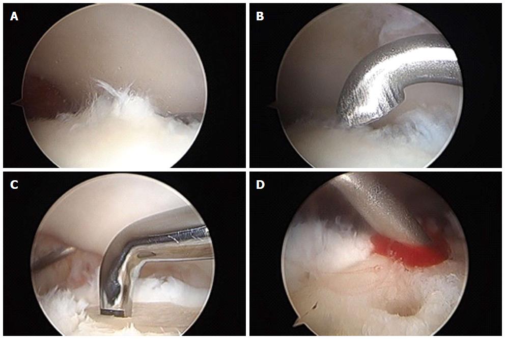
Figure 1 Arthroscopic images of osteochondral lesions of the talus.
A: Osteochondral lesion of the talus identified arthroscopically; B: Frayed or fibrillated cartilage is curretted out; C: Subchondral plate is violated with microfracture pick; D: After the subchondral bone plate is violated, bleeding occurs beginning the healing response.
- Citation: Gianakos AL, Yasui Y, Hannon CP, Kennedy JG. Current management of talar osteochondral lesions. World J Orthop 2017; 8(1): 12-20
- URL: https://www.wjgnet.com/2218-5836/full/v8/i1/12.htm
- DOI: https://dx.doi.org/10.5312/wjo.v8.i1.12


