Published online May 18, 2021. doi: 10.5312/wjo.v12.i5.254
Peer-review started: October 31, 2020
First decision: January 18, 2021
Revised: February 1, 2021
Accepted: April 5, 2021
Article in press: April 5, 2021
Published online: May 18, 2021
Processing time: 192 Days and 19.8 Hours
Isolated distal fibula fractures represent the majority of ankle fractures. These fractures are often the result of a low-energy trauma with external rotation and supination mechanism. Diagnosis is based on clinical signs and radiographic exam. Stress X-rays have a role in detecting associated mortise instability. Management depends on fracture type, displacement and associated ankle instability. For simple, minimally displaced fractures without ankle instability, conservative treatment leads to excellent results. Conservative treatment must also be considered in overaged unhealthy patients, even in unstable fractures. Surgical treatment is indicated when fracture or ankle instability are present, with several techniques described. Outcome is excellent in most cases. Complications regarding wound healing are frequent, especially with plate fixation, whereas other complications are uncommon.
Core Tip: Isolated fibula fractures are very common injuries. Diagnostic exams must rule out ankle instability. Surgical treatment must be considered in the case of associated ankle instability. Risk factors for wound related complications must be considered when choosing the surgical technique.
- Citation: Canton G, Sborgia A, Maritan G, Fattori R, Roman F, Tomic M, Morandi MM, Murena L. Fibula fractures management. World J Orthop 2021; 12(5): 254-269
- URL: https://www.wjgnet.com/2218-5836/full/v12/i5/254.htm
- DOI: https://dx.doi.org/10.5312/wjo.v12.i5.254
Ankle fractures are frequent injuries[1], increasing in elderly patients as a consequence of osteoporosis[2]. In most literature reports, distal fibula fractures represent the majority of ankle fractures[3]. These fractures are often the result of a low energy trauma with an external rotation and supination mechanism.
Many authors recommend conservative treatment for isolated fibula fractures without signs of ankle instability as good clinical results are obtained in most cases[1-3]. However, the trend in recent years is headed towards surgical treatment, with advantages in terms of anatomic restoration and earlier recovery[1-3].
Depending on fracture type, displacement and degree of instability, several surgical treatment techniques have been described. These include lateral vs posterolateral plating, nonlocking vs locking plate fixation, isolated screws and intramedullary fixation[4].
The aim of the present paper is to review the most recent literature about the epidemiology, mechanism of injury, diagnosis, classification, management and complications of isolated distal fibula fractures treatment.
Ankle fractures are frequent injuries, accounting for about 9% of all fractures[1]. Moreover, there has been a sharp increase in osteoporosis related ankle fracture incidence in recent years. Isolated distal fibula fractures represent the most frequent ankle fracture type[3,4]. Elsoe et al[2] recently reported the epidemiology of 9767 ankle fractures, identifying distal fibula fractures as the most common fracture type, accounting for 55% of cases. Furthermore, according to the Arbeitsgemeinschaft für Osteosynthesefragen/Orthopaedic Trauma Association (AO/OTA) classification, type B is generally reported to be the more common distal fibula fracture type. Court-Brown et al[5] reported the following distal fibula fracture type distribution among 1500 ankle fractures: 52% type B trans-syndesmotic fractures, 38% type A infra-syndesmotic fractures and 10% type C supra-syndesmotic fractures[5]. Age and gender-related differences in ankle fracture epidemiology have been reported. Distal fibula fractures are more frequent in young active male patients. Werner et al[6] reported an incidence of isolated distal fibula fractures reaching 83% of cases among a population of National Football League athletes reporting ankle fractures. Conversely, Hasselman et al[7] found isolated fibular fracture to cover 57.6% of cases in elderly (> 65 years) women reporting ankle fractures.
Stability of the ankle mortise is determined by bony components (fibula, tibia and talus) and ligamentous structures (syndesmosis complex and lateral and medial collateral ligaments). Dynamic musculotendinous stabilizers, which exact function is less understood, also play a relevant role. There is on-going research on interactions between these structures and mechanisms that cause fracture.
Ankle sprains/torsion injuries, accidental falls and sports related accidents are the most frequently reported causes of distal fibula fracture, with different rates according to the different AO/OTA types. In type A fractures the main cause is represented by torsion (32%) followed by falls (23%) and sports related trauma (22%). In type B fractures the trend is similar, with reported rates of 27% for torsion, 37% for falls and 13% for sports related trauma. For type C the trend is slightly different because torsion represents only 3.7% of cases while falls and sports related trauma represent 28% and 21% of cases, respectively.
The most frequently described traumatic mechanism is supination-external rotation (SER). In this type of trauma, the talus rotates pushing the tibia and fibula apart, rupturing the anterior-inferior tibiofibular ligament and causing a simple ankle sprain. Further progression of deforming force causes a simple oblique fibula fracture at the level of the syndesmosis, equivalent to the AO/OTA type B fracture. In the second most frequent mechanism, supination-adduction, adduction of the hindfoot causes either talofibular ligament rupture (ankle sprain) or an avulsion fracture of the distal fibula, equivalent to the AO/OTA type A fracture. As reported by Lauge-Hansen[8], lateral structures are damaged after the medial side when traumatic forces act in pronation.
Nonetheless, a recent in vivo study by Kwon et al[9] analyzing injury videos posted on YouTube and matching them to their corresponding X-rays, found that the Lauge-Hansen system was only 58% overall accurate in predicting fracture patterns from deforming injury mechanism.
Clinical signs of an isolated fracture of the distal fibula are not specific and may not be distinguishable from a severe ankle sprain. These signs include swelling, bruising, pain, ecchymosis, tenderness and reduced range of motion (ROM). Swelling is the most common reported sign and was found to be a constant feature of all ankle fractures[10]. Because most isolated distal fibula fractures are stable, weight bearing is usually possible[11], thus patients might be ambulating at clinical presentation. There are no specific clinical tests for this fracture. Nonetheless, it is essential to evaluate medial ankle structures stability to choose the correct management. In C type fractures, the syndesmosis complex integrity must also be investigated. However, clinical examination alone is not diagnostic in most cases because pain, edema and muscle contracture can hinder correct evaluation[12,13].
Standard ankle X-rays are the mainstay of instrumental diagnosis for all ankle fractures. However, ankle sprains that might possibly cause a fracture might not deserve radiographic examination in all cases. In fact, due to the already described unspecific clinical presentation, other criteria should be considered to reduce the number of unnecessary exams and length of hospital stay[14,15]. The Ottawa Criteria were introduced for this purpose, despite some studies questioning their clinical validity[16].
When cases amenable for radiographic evaluation are selected, three radiographic views should always be obtained according to the American College of Radiology guidelines: antero-posterior (AP), lateral and mortise view[17].
The AP view is performed along the long axis of the foot. In isolated fibula fractures, this view is particularly useful to evaluate signs of associated ankle and/or syndes
In the lateral view, the talar dome must be centered and congruent with the tibial plafond. This view is useful in isolated fibula fractures to demonstrate AP displace
The mortise view is taken by placing the foot on the table with about 15° of internal rotation. This visualization is useful in isolated fibula fractures to detect signs of associated syndesmosis instability and to obtain a clear view of the lateral malleolus without other overlapping structures. It is especially useful in undisplaced and incomplete fractures[18,20].
Theoretically, these views are sufficient to identify an isolated distal fibula fracture in almost all cases. As demonstrated in several clinical and cadaveric studies[21-23] in standard radiographic evaluation, a MCS increase in isolated distal fibula fractures is a typical sign of rupture of the deltoid ligament with consequent talar lateral shift. These factors suggest a possible mortise instability, which is fundamental to define treatment modality. However, reliable radiographic determination of deltoid ligament rupture in such uncertain cases is difficult in clinical practice[24]. To adequately evaluate mortise stability, different modalities to obtain stress radiographs have been described.
The manual stress view has been considered the method of choice for years. However, being an operator dependent exam, reproducibility and radiation exposure to the physician are a concern. Moreover, it is not clear in the published data which MCS values are to be considered as a cut off to identify a clinically relevant deltoid ligament rupture.
Michelson et al[25] described for the first time in 2001 the gravity stress view to investigate ankle joint instability. The patient is placed in the lateral decubitus position with the distal half of the leg off the end of the table. Then a standard AP and mortise view is taken. Gravity stress views proved to be just as effective as manual stress views to detect deltoid ligament injury in association with an isolated distal fibula fracture[26-28]. Many advantages, such as no radiation exposure to the physician and less pain to the patient are described. Moreover, the constant force of gravity makes the exam reproducible[27].
Nonetheless, manual and gravity stress radiographs can overestimate the need for surgical fixation by showing MCS widening in partial deltoid ligament lesions that might uneventfully heal with conservative treatment[29-31]. For this reason, in the literature many authors described the weight bearing radiographs as an alternative method to identify ankle instability in isolated distal fibula fractures. As proposed by Weber et al[32], these radiographs should be performed with the patient standing on both legs as pain allows. An AP, lateral and mortise view is then taken with the weight ideally distributed equally on both ankles[32]. The disadvantage of this technique is the possible variability in weight distribution between healthy and injured side that could hide the degree of ankle instability in some cases.
Finally, magnetic resonance imaging is not indicated in the acute diagnosis of isolated distal fibula fractures. Its use in these lesions is very limited because traditional and stress radiographs proved to be equally effective in identifying the severity of associated deltoid ligament injury[33]. Conversely, magnetic resonance imaging might be useful to detect associated chondral injuries or to diagnose a fracture when conventional radiographs are inconclusive, and clinical suspicion is very high[34].
Although the fibula carries only 10% of the body weight (compared to 90% carried by the tibia)[35], its role is crucial in the stability of the ankle mortise. Based on this statement, to define an ankle with isolated fibula fracture as stable or unstable is crucial to guide proper treatment. Nonetheless, there is still debate in the literature on which are the most suitable clinical and radiological criteria to obtain this goal. The optimal management is based on an accurate knowledge of the fracture. For a comprehensive assessment of the fracture, a reproducible classification method is essential.
Different classifications have been proposed through the years. Lauge-Hansen and Danis-Weber classifications are the most used. They are based on standard AP, lateral and mortise radiographic views of the ankle. Their aim is to describe the mechanism of injury, predict soft tissue conditions and finally guide treatment.
The Lauge-Hansen classification, developed in 1954, is based on the position of the foot at the time of injury (supination or pronation) and on the deforming forces acting on the foot (abduction, adduction or external rotation)[36]. This results in a combi
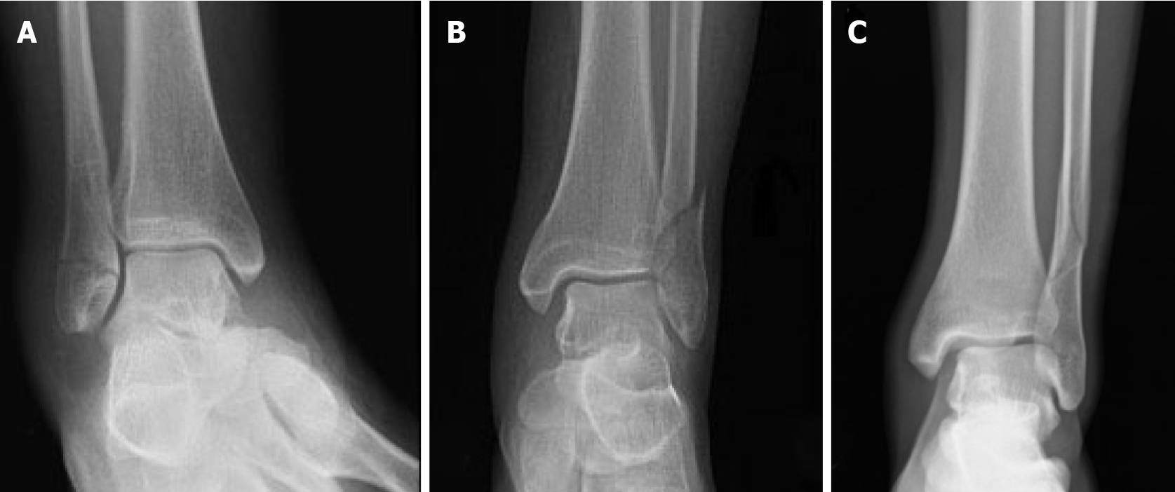
The Danis-Weber classification (Figure 1) was first described by Robert Danis in 1949 and later modified by Bernhard Georg Weber in 1966. It was then adopted by the AO/OTA Group. It evaluates the location of the main fibular fracture in relation to the syndesmosis. Type A fractures are generally stable injuries occurring below the level of the syndesmosis. Type B fractures occur at the level of the syndesmosis and might be unstable in some cases. Type C fractures are usually unstable injuries occurring above the level of the syndesmosis[37].
However, differentiating fracture types in relation to the syndesmosis might lead the medial side of the ankle to be overlooked. Moreover, injury extent in the tibiofibular syndesmosis is often not predictable. These findings are crucial to detect tibio-talar instability and consequently to decide between surgical and nonsurgical management, especially for type B fractures[38]. Probably, as suggested by Lampridis et al[39], a combination of the two main systems is the correct approach. Nonetheless, the limits of these classification systems are reported by several studies in the literature, especially their poor prognostic and therapeutically predictive capabili
Clinical and biomechanical data indicate that maintenance of talar reduction is the most important factor for the prognosis of ankle fractures[43,44]. A residual disloca
Shortening and external rotation of the fibula can cause talar lateral shift, reducing the tibio-talar contact area and increasing peak pressure in the articular cartila
An isolated distal fibula fracture is considered stable when less than 2 mm of displacement occurs, and no deltoid ligament rupture is detected (MCS < 4 mm)[50,51]. Several clinical studies have shown that in isolated fractures without concomitant medial injury, conservative treatment leads to excellent long-term results[52,53].
Conversely, many clinical studies have shown significantly better results in ankle fractures with mortise instability and talus displacement when an accurate fracture reduction is achieved[54]. However, unlike cases associated with gross instability, proper management of isolated fibula fractures that demonstrate instability only after stress radiographs is still a matter of debate in the literature[55]. In many authors’ opinions, stress radiographs can overestimate the need for surgical fixation[29-31]. Hoshino et al[31] analyzed 36 patients with isolated distal fibula fracture demon
Conservative treatment is indicated for isolated distal fibula fractures with a stable ankle mortise. As far as fracture displacement is concerned, Lesic et al[56] set 2 mm as the threshold between conservative and surgical treatment. However, there is no strong evidence in the literature advocating surgery for fracture displacement more than 2 mm[57]. Other studies suggest that radiographic displacement might not be reliable as it is mostly a rotational displacement[58-60].
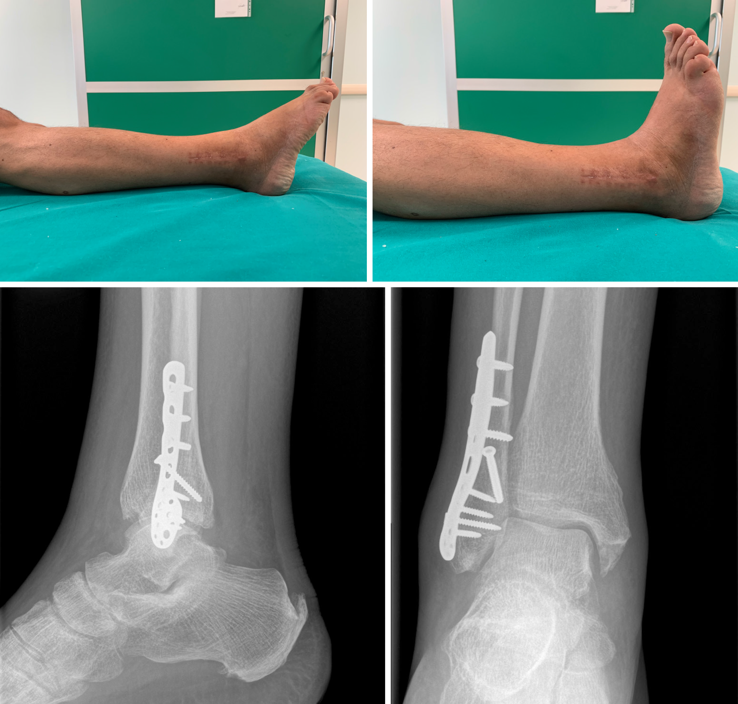
Nonetheless, minor radiographic displacement seems not to affect clinical outcome[61]. Two studies have shown a high percentage of good results even when the fibula is posteriorly displaced up to 5 mm[41,62]. Hence, any isolated distal fibula fracture with a stable ankle can be treated conservatively (Figure 2).
Weber type A fractures can be considered equivalent to a ligamentous ankle injury. Likely, satisfactory results with nonoperative treatment can be achieved as in ligament ruptures[63].
In the literature, randomized and nonrandomized studies show satisfactory outcomes for conservative treatment in minimally displaced or nondisplaced Weber B type fractures[41,61,62,64]. Dobbe et al[65] reported that 13% of 108 conservatively treated infra-syndesmotic fractures had difficulties with work- and life-related activities. However, no relationship was identified between outcomes and the degree of articular displacement or fragment width[65]. These results might suggest the need to adapt treatment according to age and activity level. Sanders et al[54] suggested operative intervention in younger individuals, with the aim to reduce the risk of malalignment and improve outcomes. Conversely, a different management can be reserved for older and less active patients, which can be safely treated with cast immobilization even in unstable fracture patterns[54].
While there are several studies that describe conservative treatment for Weber type B fractures, a recent review comparing different managements for Weber type C fractures found only one study included conservative treatment. This demonstrates the widespread preference for surgical management in these cases[66]. Donken et al[67] compared the results of nonoperative and operative treatment for Weber type C fractures, with conservative treatment reserved to cases that demonstrated joint congruity, no signs of deltoid ligament injury and no medial malleolus fractures. Clinical results were comparable with most patients reaching high-level functional results[67].
Nonoperative treatment modality is chosen based on patients’ symptoms, bone and skin quality, time lapse from injury to clinical presentation and risk factors for impaired healing (Table 1). Different conservative treatment modalities are described in the literature, ranging from cast immobilization without weight bearing to immediate full weight bearing without cast or brace. Historically, treatment in a plaster cast for several weeks was recommended. This strategy arises from the evidence that 6 wk are needed for any fracture to tolerate weight bearing[41,61,62,68]. Although a high rate of fracture union was demonstrated, a prolonged immobilization can result in ankle stiffness and higher risk of deep vein thrombosis[69-73]. To overcome these complications, Kortekangas et al[74] recently showed that a 3-wk period of immobilization is noninferior to 6 wk in the treatment of an isolated stable Weber B type fracture.
| Nonoperative treatment | |
| Displacement | < 2 mm |
| Medial stability | MCS < 4 mm in AP/mortise view and/or in dynamic radiographs view |
| Poor bone and skin quality | |
| Long time lapse from injury | |
| Advanced age, low functional demand | |
| High risk of local and general complications | |
Alternative methods of immobilization with immediate weight bearing have been proposed through the years. In 1979, Stover et al[75] proposed a bivalve pneumatic air stirrup in the management of ankle fractures. Stuart et al[71] in 1989 showed the brace to improve patient comfort, post fracture swelling, range of ankle motion at union and time to full rehabilitation. Similar findings were reported by Brink et al[76], who advocated the use of a hinged short-leg boot with good results in pain relief, increased ROM and earlier return to ambulation. It has been suggested that an ordinary elastic bandage is equally safe and beneficial[52], and no difference in the amount of pain experienced has been found between early mobilization or plaster cast[77]. Ryd et al[52] described 49 patients treated only with elastic bandage for isolated distal fibula fractures displaced less than 2 mm and without medial tenderness. All of them had excellent clinical outcomes and were back to normal activity in about 4 mo[52]. The functional treatment of stable ankle fractures is also supported by van der Berg et al[78], who showed better Visual Analogue Scale score and total ROM with a brace rather than with a cast after 6 wk, while no significant difference was found at 1 year.
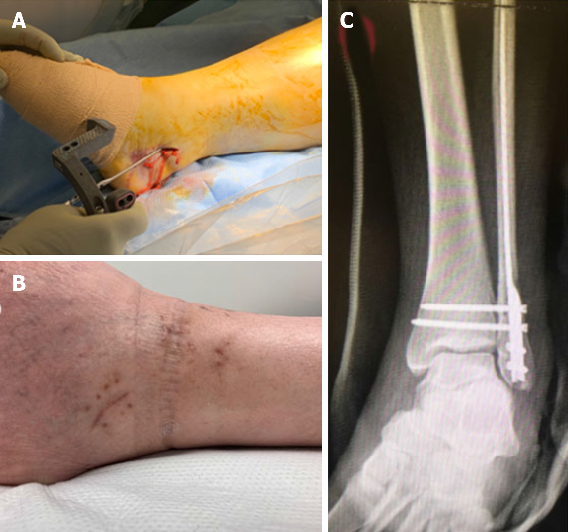
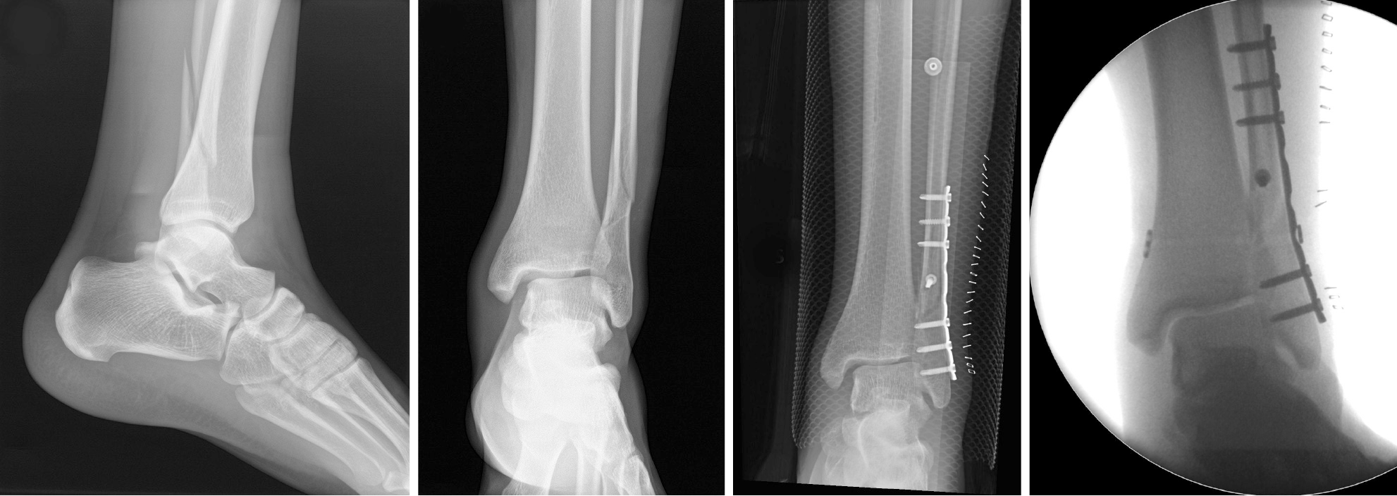
Open reduction and internal fixation is the most common treatment for unstable ankle fractures (Table 2). There are several fixation methods described for distal fibula fractures fixation, including one-third tubular plate (Figure 3), dynamic compression plate and locking plate with or without an independent lag screw[79-81]. The most used plates are angular stable metaphyseal or anatomic distal fibula plates (Figure 4)[82]. They can serve as bridging plates, compression plates, tension band plates or neutralization plates. Most studies comparing locking plates and conventional one third tubular plates show no differences in clinical and radiographic outcomes as well as in wound complications incidence[79,83-85]. However, these fixation techniques have a complication rate of up to 30% of cases, with wound complications being the most common[86-88]. This is attributed to the surgical trauma occurring in an already injured area with limited soft tissue cover. This range increases in smokers, in elderly patients and in patients with comorbidities such as diabetes and peripheral vascu
| Surgical treatment | |
| Displacement | > 2-5 mm |
| Medial instability | MCS > 4 mm in AP/mortise view or in dynamic radiographs view |
| Type C | Any displacement |
Distal fibula fractures in elderly patients are often comminuted and present with impaired soft tissues coverage. Consequently, the correct management of ankle fractures in these patients must account for bone quality and the risk of soft tissue complications[80,86,89]. Locking plates provide a biomechanical advantage in cases of poor bone quality and are therefore recommended over nonlocking plates in osteoporotic patients when surgical management is chosen[86,80].
Fibular nailing is considered a valid alternative method of fixation for distal fibula fractures. The use of intramedullary fibula fixation was first introduced in the mid-1980s to reduce complications of the traditional plating techniques[91]. However, early attempts in intramedullary fibula fixation, such as using rush rods, Inyo nail, K. wires, etc., showed several complications and failures due to poor rotational and longitudinal stability, which led to loss of reduction, malunion and nail migration[92-94]. As a result, modern locking fibula nails have been developed to reduce such complica
Nonunion in ankle fractures is an extremely rare complication[99-103]. Distal fibula fractures usually heal uneventfully even with nonsurgical treatment. However, because most nonunions in this area are asymptomatic, the exact incidence of this complication is uncertain[99-103].
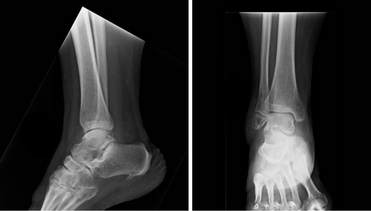
A higher incidence of fibular nonunion has been described when associated with medial instability. Sneppen et al[99] demonstrated a 0.7% incidence of fibular nonunion in ankle fractures involving the medial malleolus vs a 0.1% incidence in isolated fibular fractures. Complete nonunion was mostly seen in type A (Figure 6) and C fractures[100], whereas incomplete nonunion was described in Weber type B fractures[101].
Treatment is based on the type of nonunion, symptoms, initial fracture pattern, associated injuries and patient expectations. Asymptomatic nonunions are treated conservatively, with reports of spontaneous healing even several years after the initial injury[102]. For symptomatic nonunion, open reduction and internal fixation with or without bone grafting represents the best treatment choice resulting in successful outcomes in most cases[103].
Angular malalignment occurs when the distal fibula heals in shortening or external rotation. This causes a lateral subluxation of the talus with ankle kinematics alteration leading to arthritis[44]. Most cases of angular malalignment occur after conservative treatment[104]. However, surgical treatment might be a cause of malunion if anatomic reduction is not achieved intraoperatively. Moreover, technical errors, suboptimal stability of fixation and unrecognized associated ligamentous instability might lead to loss of reduction and consequent malunion (Figure 7)[105]. Several radiographic parameters have been described to identify the correct length and rotation of the fibula in the ankle mortise: the Weber circle, the Shenton’s line, the talocrural and bimalleolar angles[106]. These parameters can help the clinicians to more easily identify a malalignment that could cause continuous pain to the patient even after several months.
Surgical treatment is a demanding procedure, as anatomic reconstruction usually requires both fibula osteotomy and soft tissues release. Plate osteosynthesis is then required for fixation[107]. In several studies, the results of surgical treatment for malalignment were excellent. Yablon et al[108] reported good results over 7 years of follow up in 23 of 26 patients with fibular malalignment surgically treated 6 years after trauma. Ward et al[109] reported similar results in ankle fracture malunions treated with lengthening osteotomy of the fibula.
The distal fibula is subcutaneous and lacks a layer of overlying muscles. Thus, wound healing complications are the most common adverse events related to distal fibula fixation. Wound edge necrosis, wound dehiscence and superficial and deep infection have all been reported[110]. In the literature, the overall wound complication rate varies from 8.4% to 40.0% among studies[111-113] . The deep surgical infection rate is significantly lower, about 1.2% to 2.8% according to different reports[111,114,115].
The genesis is multifactorial as it depends on soft tissue compromise at admission, timing of surgery, fracture type and patient characteristics. Age, diabetes, steroid intake, smoking and peripheral vascular disease are associated with a greater risk of wound complications[88,112,116-118].The type of implant also might have a role. Schepers et al[119] reported in a retrospective study of 165 patients a higher wound complication rate with locking plates (17.5%) than with thinner one third tubular plates (5.5%). Conversely, Tsukada et al[84] in two different trials did not find any difference in complication rates between locking and nonlocking plates[84,115].
The incidence of this complication can be minimized by treating the fracture as soon as reasonably possible[110] or postponing the surgery until the edema is resolved. The latter strategy has gained more and more popularity with the advent of damage control techniques. Whatever the timing, limiting the use of the tourniquet and closing the wound without tension is also advisable.
Most patients after isolated distal fibula fractures recover with a completely functional ROM. When stiffness occurs, dorsiflexion deficit is more common. Lin et al[120] reported that in a population of 306 patients a 19% rate of plantar flexion limitation (only 2% > 10 degrees) occurred, while 41% of cases had restriction in dorsiflexion. In a review of 31 randomized trials about rehabilitation of ankle fractures, the authors found a positive effect on ankle ROM form early mobilization, early weight bearing and the use of a removable immobilization device. However, there is limited evidence supporting this strategy as patient compliance seems to play a significant role[120].
Trauma is the most common cause of ankle osteoarthritis[121]. Arthritis results from a combination of direct cartilage damage and biomechanical alterations that affect joint kinematics[122]. It becomes evident 2 to 3 years after trauma and is often symptomatic. It is more frequent in higher grade fractures according to the Lauge-Hansen classification. Lübbeke et al[105], evaluating risk factors for development of ankle osteo
Isolated fibula fractures are very common injuries. Diagnostic exams must rule out ankle instability. Conservative treatment yields good results in stable fractures with stable ankle mortise. Open reduction internal fixation is indicated in case of associated ankle instability. Risk factors for wound related complications must be considered when choosing a surgical technique.
Manuscript source: Invited manuscript
Specialty type: Orthopedics
Country/Territory of origin: Italy
Peer-review report’s scientific quality classification
Grade A (Excellent): 0
Grade B (Very good): B
Grade C (Good): 0
Grade D (Fair): D
Grade E (Poor): 0
P-Reviewer: Amornyotin S, Tsikopoulos K S-Editor: Liu M L-Editor: Filipodia P-Editor: Yuan YY
| 1. | Court-Brown CM, Caesar B. Epidemiology of adult fractures: A review. Injury. 2006;37:691-697. [RCA] [PubMed] [DOI] [Full Text] [Cited by in Crossref: 1916] [Cited by in RCA: 2185] [Article Influence: 115.0] [Reference Citation Analysis (0)] |
| 2. | Elsoe R, Ostgaard SE, Larsen P. Population-based epidemiology of 9767 ankle fractures. Foot Ankle Surg. 2018;24:34-39. [RCA] [PubMed] [DOI] [Full Text] [Cited by in Crossref: 135] [Cited by in RCA: 202] [Article Influence: 28.9] [Reference Citation Analysis (1)] |
| 3. | Jehlicka D, Bartonícek J, Svatos F, Dobiás J. [Fracture-dislocations of the ankle joint in adults. Part I: epidemiologic evaluation of patients during a 1-year period]. Acta Chir Orthop Traumatol Cech. 2002;69:243-247. [PubMed] |
| 4. | Aiyer AA, Zachwieja EC, Lawrie CM, Kaplan JRM. Management of Isolated Lateral Malleolus Fractures. J Am Acad Orthop Surg. 2019;27:50-59. [RCA] [PubMed] [DOI] [Full Text] [Cited by in Crossref: 21] [Cited by in RCA: 34] [Article Influence: 5.7] [Reference Citation Analysis (1)] |
| 5. | Court-Brown CM, McBirnie J, Wilson G. Adult ankle fractures--an increasing problem? Acta Orthop Scand. 1998;69:43-47. [RCA] [PubMed] [DOI] [Full Text] [Cited by in Crossref: 425] [Cited by in RCA: 449] [Article Influence: 16.6] [Reference Citation Analysis (0)] |
| 6. | Werner BC, Mack C, Franke K, Barnes RP, Warren RF, Rodeo SA. Distal Fibula Fractures in National Football League Athletes. Orthop J Sports Med. 2017;5:2325967117726515. [RCA] [PubMed] [DOI] [Full Text] [Full Text (PDF)] [Cited by in Crossref: 7] [Cited by in RCA: 7] [Article Influence: 0.9] [Reference Citation Analysis (0)] |
| 7. | Hasselman CT, Vogt MT, Stone KL, Cauley JA, Conti SF. Foot and ankle fractures in elderly white women. Incidence and risk factors. J Bone Joint Surg Am. 2003;85:820-824. [RCA] [PubMed] [DOI] [Full Text] [Cited by in Crossref: 155] [Cited by in RCA: 154] [Article Influence: 7.0] [Reference Citation Analysis (1)] |
| 8. | Lauge-Hansen N. Fractures of the ankle. II. Combined experimental-surgical and experimental-roentgenologic investigations. Arch Surg. 1950;60:957-985. [RCA] [PubMed] [DOI] [Full Text] [Cited by in Crossref: 659] [Cited by in RCA: 568] [Article Influence: 27.0] [Reference Citation Analysis (0)] |
| 9. | Kwon JY, Chacko AT, Kadzielski JJ, Appleton PT, Rodriguez EK. A novel methodology for the study of injury mechanism: ankle fracture analysis using injury videos posted on YouTube.com. J Orthop Trauma. 2010;24:477-482. [RCA] [PubMed] [DOI] [Full Text] [Cited by in Crossref: 26] [Cited by in RCA: 30] [Article Influence: 2.0] [Reference Citation Analysis (0)] |
| 10. | Montague AP, McQuillan RF. Clinical assessment of apparently sprained ankle and detection of fracture. Injury. 1985;16:545-546. [RCA] [PubMed] [DOI] [Full Text] [Cited by in Crossref: 21] [Cited by in RCA: 20] [Article Influence: 0.5] [Reference Citation Analysis (0)] |
| 11. | Lynch SA. Assessment of the Injured Ankle in the Athlete. J Athl Train. 2002;37:406-412. [PubMed] |
| 12. | Gebing R, Fiedler V. [X-ray diagnosis of ligament lesions of the superior ankle joint]. Radiologe. 1991;31:594-600. [PubMed] |
| 13. | Oloff LM, Sullivan BT, Heard GS, Thornton MC. Magnetic resonance imaging of traumatized ligaments of the ankle. J Am Podiatr Med Assoc. 1992;82:25-32. [RCA] [PubMed] [DOI] [Full Text] [Cited by in Crossref: 15] [Cited by in RCA: 14] [Article Influence: 0.4] [Reference Citation Analysis (1)] |
| 14. | Stiell IG, Greenberg GH, McKnight RD, Nair RC, McDowell I, Reardon M, Stewart JP, Maloney J. Decision rules for the use of radiography in acute ankle injuries. Refinement and prospective validation. JAMA. 1993;269:1127-1132. [RCA] [PubMed] [DOI] [Full Text] [Cited by in Crossref: 344] [Cited by in RCA: 299] [Article Influence: 9.3] [Reference Citation Analysis (0)] |
| 15. | Stiell I, Wells G, Laupacis A, Brison R, Verbeek R, Vandemheen K, Naylor CD. Multicentre trial to introduce the Ottawa ankle rules for use of radiography in acute ankle injuries. Multicentre Ankle Rule Study Group. BMJ. 1995;311:594-597. [RCA] [PubMed] [DOI] [Full Text] [Cited by in Crossref: 180] [Cited by in RCA: 161] [Article Influence: 5.4] [Reference Citation Analysis (0)] |
| 16. | Stiell IG, Greenberg GH, McKnight RD, Wells GA. The "real" Ottawa ankle rules. Ann Emerg Med. 1996;27:103-104. [RCA] [PubMed] [DOI] [Full Text] [Cited by in Crossref: 17] [Cited by in RCA: 16] [Article Influence: 0.6] [Reference Citation Analysis (1)] |
| 17. | Dalinka MK, Alazraki N, Berquist TH, Daffner RH, DeSmet AA, el-Khoury GY, Goergen TG, Keats TE, Manaster BJ, Newberg A, Pavlov H, Haralson RH 3rd, McCabe JB, Sartoris D. Imaging evaluation of suspected ankle fractures. American College of Radiology. ACR Appropriateness Criteria. Radiology. 2000;215 Suppl:239-241. [PubMed] |
| 18. | Krähenbühl N, Weinberg MW, Davidson NP, Mills MK, Hintermann B, Saltzman CL, Barg A. Imaging in syndesmotic injury: a systematic literature review. Skeletal Radiol. 2018;47:631-648. [RCA] [PubMed] [DOI] [Full Text] [Cited by in Crossref: 59] [Cited by in RCA: 74] [Article Influence: 10.6] [Reference Citation Analysis (0)] |
| 19. | Lampignano JP, Kendrick LE. Bontrager’s Textbook of Radiographic Positioning and Related Anatomy. 9th ed. Mosby, 2017: 239-242. |
| 20. | Patel P, Russell TG. Ankle Radiographic Evaluation. [cited 14 September 2020]. In: StatPearls [Internet]. Treasure Island (FL): StatPearls. [PubMed] |
| 21. | Harper MC. The deltoid ligament. An evaluation of need for surgical repair. Clin Orthop Relat Res. 1988;(226):156-168. [RCA] [PubMed] [DOI] [Full Text] [Cited by in Crossref: 51] [Cited by in RCA: 46] [Article Influence: 1.2] [Reference Citation Analysis (0)] |
| 22. | Harper MC. Deltoid ligament: an anatomical evaluation of function. Foot Ankle. 1987;8:19-22. [RCA] [PubMed] [DOI] [Full Text] [Cited by in Crossref: 130] [Cited by in RCA: 117] [Article Influence: 3.1] [Reference Citation Analysis (0)] |
| 23. | Baird RA, Jackson ST. Fractures of the distal part of the fibula with associated disruption of the deltoid ligament. Treatment without repair of the deltoid ligament. J Bone Joint Surg Am. 1987;69:1346-1352. [RCA] [PubMed] [DOI] [Full Text] [Cited by in Crossref: 106] [Cited by in RCA: 96] [Article Influence: 2.6] [Reference Citation Analysis (0)] |
| 24. | Schuberth JM, Collman DR, Rush SM, Ford LA. Deltoid ligament integrity in lateral malleolar fractures: a comparative analysis of arthroscopic and radiographic assessments. J Foot Ankle Surg. 2004;43:20-29. [RCA] [PubMed] [DOI] [Full Text] [Cited by in Crossref: 93] [Cited by in RCA: 89] [Article Influence: 4.2] [Reference Citation Analysis (0)] |
| 25. | Michelson JD, Varner KE, Checcone M. Diagnosing deltoid injury in ankle fractures: the gravity stress view. Clin Orthop Relat Res. 2001;(387):178-182. [RCA] [PubMed] [DOI] [Full Text] [Cited by in Crossref: 108] [Cited by in RCA: 108] [Article Influence: 4.5] [Reference Citation Analysis (0)] |
| 26. | Gill JB, Risko T, Raducan V, Grimes JS, Schutt RC Jr. Comparison of manual and gravity stress radiographs for the evaluation of supination-external rotation fibular fractures. J Bone Joint Surg Am. 2007;89:994-999. [RCA] [PubMed] [DOI] [Full Text] [Cited by in Crossref: 78] [Cited by in RCA: 82] [Article Influence: 4.6] [Reference Citation Analysis (0)] |
| 27. | Schock HJ, Pinzur M, Manion L, Stover M. The use of gravity or manual-stress radiographs in the assessment of supination-external rotation fractures of the ankle. J Bone Joint Surg Br. 2007;89:1055-1059. [RCA] [PubMed] [DOI] [Full Text] [Cited by in Crossref: 92] [Cited by in RCA: 96] [Article Influence: 5.3] [Reference Citation Analysis (0)] |
| 28. | LeBa TB, Gugala Z, Morris RP, Panchbhavi VK. Gravity vs manual external rotation stress view in evaluating ankle stability: a prospective study. Foot Ankle Spec. 2015;8:175-179. [RCA] [PubMed] [DOI] [Full Text] [Cited by in Crossref: 23] [Cited by in RCA: 27] [Article Influence: 2.7] [Reference Citation Analysis (0)] |
| 29. | Dawe EJ, Shafafy R, Quayle J, Gougoulias N, Wee A, Sakellariou A. The effect of different methods of stability assessment on fixation rate and complications in supination external rotation (SER) 2/4 ankle fractures. Foot Ankle Surg. 2015;21:86-90. [RCA] [PubMed] [DOI] [Full Text] [Cited by in Crossref: 24] [Cited by in RCA: 27] [Article Influence: 2.7] [Reference Citation Analysis (0)] |
| 30. | Hastie GR, Akhtar S, Butt U, Baumann A, Barrie JL. Weightbearing Radiographs Facilitate Functional Treatment of Ankle Fractures of Uncertain Stability. J Foot Ankle Surg. 2015;54:1042-1046. [RCA] [PubMed] [DOI] [Full Text] [Cited by in Crossref: 28] [Cited by in RCA: 29] [Article Influence: 2.9] [Reference Citation Analysis (0)] |
| 31. | Hoshino CM, Nomoto EK, Norheim EP, Harris TG. Correlation of weightbearing radiographs and stability of stress positive ankle fractures. Foot Ankle Int. 2012;33:92-98. [RCA] [PubMed] [DOI] [Full Text] [Cited by in Crossref: 54] [Cited by in RCA: 60] [Article Influence: 4.6] [Reference Citation Analysis (1)] |
| 32. | Weber M, Burmeister H, Flueckiger G, Krause FG. The use of weightbearing radiographs to assess the stability of supination-external rotation fractures of the ankle. Arch Orthop Trauma Surg. 2010;130:693-698. [RCA] [PubMed] [DOI] [Full Text] [Cited by in Crossref: 74] [Cited by in RCA: 78] [Article Influence: 5.2] [Reference Citation Analysis (0)] |
| 33. | Nortunen S, Lepojärvi S, Savola O, Niinimäki J, Ohtonen P, Flinkkilä T, Lantto I, Kortekangas T, Pakarinen H. Stability assessment of the ankle mortise in supination-external rotation-type ankle fractures: lack of additional diagnostic value of MRI. J Bone Joint Surg Am. 2014;96:1855-1862. [RCA] [PubMed] [DOI] [Full Text] [Cited by in Crossref: 54] [Cited by in RCA: 59] [Article Influence: 5.4] [Reference Citation Analysis (0)] |
| 34. | Sadineni RT, Pasumarthy A, Bellapa NC, Velicheti S. Imaging Patterns in MRI in Recent Bone Injuries Following Negative or Inconclusive Plain Radiographs. J Clin Diagn Res. 2015;9:TC10-TC13. [RCA] [PubMed] [DOI] [Full Text] [Cited by in Crossref: 5] [Cited by in RCA: 11] [Article Influence: 1.1] [Reference Citation Analysis (0)] |
| 35. | Crist B, Fischer SJ, Dunbar RP. Ankle Fractures (Broken Ankle). [cited 27 August 2020]. In: American Academy of Orthopedic Surgeons [Internet]. Last reviewed: March 2013. Available from: https://orthoinfo.aaos.org/en/diseases--conditions/ankle-fractures-broken-ankle/. |
| 36. | Lauge-Hansen N. Fractures of the ankle. III. Genetic roentgenologic diagnosis of fractures of the ankle. Am J Roentgenol Radium Ther Nucl Med. 1954;71:456-471. [PubMed] |
| 37. | Danis R. Les fractures malleolaires. In: Danis R. Théorie et pratique de l’ostéosynthèse. Paris: Masson, 1949: 133-165. |
| 38. | Fonseca LLD, Nunes IG, Nogueira RR, Martins GEV, Mesencio AC, Kobata SI. Reproducibility of the Lauge-Hansen, Danis-Weber, and AO classifications for ankle fractures. Rev Bras Ortop. 2018;53:101-106. [RCA] [PubMed] [DOI] [Full Text] [Full Text (PDF)] [Cited by in Crossref: 4] [Cited by in RCA: 22] [Article Influence: 2.8] [Reference Citation Analysis (1)] |
| 39. | Lampridis V, Gougoulias N, Sakellariou A. Stability in ankle fractures: Diagnosis and treatment. EFORT Open Rev. 2018;3:294-303. [RCA] [PubMed] [DOI] [Full Text] [Full Text (PDF)] [Cited by in Crossref: 52] [Cited by in RCA: 56] [Article Influence: 8.0] [Reference Citation Analysis (0)] |
| 40. | Broos PL, Bisschop AP. Operative treatment of ankle fractures in adults: correlation between types of fracture and final results. Injury. 1991;22:403-406. [RCA] [PubMed] [DOI] [Full Text] [Cited by in Crossref: 141] [Cited by in RCA: 137] [Article Influence: 4.0] [Reference Citation Analysis (0)] |
| 41. | Bauer M, Jonsson K, Nilsson B. Thirty-year follow-up of ankle fractures. Acta Orthop Scand. 1985;56:103-106. [RCA] [PubMed] [DOI] [Full Text] [Cited by in Crossref: 176] [Cited by in RCA: 151] [Article Influence: 3.8] [Reference Citation Analysis (0)] |
| 42. | Egol KA, Amirtharajah M, Tejwani NC, Capla EL, Koval KJ. Ankle stress test for predicting the need for surgical fixation of isolated fibular fractures. J Bone Joint Surg Am. 2004;86:2393-2398. [RCA] [PubMed] [DOI] [Full Text] [Cited by in Crossref: 128] [Cited by in RCA: 120] [Article Influence: 5.7] [Reference Citation Analysis (0)] |
| 43. | Michelsen JD, Ahn UM, Helgemo SL. Motion of the ankle in a simulated supination-external rotation fracture model. J Bone Joint Surg Am. 1996;78:1024-1031. [RCA] [PubMed] [DOI] [Full Text] [Cited by in Crossref: 107] [Cited by in RCA: 92] [Article Influence: 3.2] [Reference Citation Analysis (0)] |
| 44. | Clarke HJ, Michelson JD, Cox QG, Jinnah RH. Tibio-talar stability in bimalleolar ankle fractures: a dynamic in vitro contact area study. Foot Ankle. 1991;11:222-227. [RCA] [PubMed] [DOI] [Full Text] [Cited by in Crossref: 83] [Cited by in RCA: 74] [Article Influence: 2.2] [Reference Citation Analysis (0)] |
| 45. | Curtis MJ, Michelson JD, Urquhart MW, Byank RP, Jinnah RH. Tibiotalar contact and fibular malunion in ankle fractures. A cadaver study. Acta Orthop Scand. 1992;63:326-329. [RCA] [PubMed] [DOI] [Full Text] [Cited by in Crossref: 65] [Cited by in RCA: 67] [Article Influence: 2.0] [Reference Citation Analysis (1)] |
| 46. | Kimizuka M, Kurosawa H, Fukubayashi T. Load-bearing pattern of the ankle joint. Contact area and pressure distribution. Arch Orthop Trauma Surg. 1980;96:45-49. [RCA] [PubMed] [DOI] [Full Text] [Cited by in Crossref: 116] [Cited by in RCA: 111] [Article Influence: 2.5] [Reference Citation Analysis (0)] |
| 47. | Moody ML, Koeneman J, Hettinger E, Karpman RR. The effects of fibular and talar displacement on joint contact areas about the ankle. Orthop Rev. 1992;21:741-744. [PubMed] |
| 48. | Michelson JD, Clarke HJ, Jinnah RH. The effect of loading on tibiotalar alignment in cadaver ankles. Foot Ankle. 1990;10:280-284. [RCA] [PubMed] [DOI] [Full Text] [Cited by in Crossref: 57] [Cited by in RCA: 52] [Article Influence: 1.5] [Reference Citation Analysis (0)] |
| 49. | Tornetta P 3rd. Competence of the deltoid ligament in bimalleolar ankle fractures after medial malleolar fixation. J Bone Joint Surg Am. 2000;82:843-848. [RCA] [PubMed] [DOI] [Full Text] [Cited by in Crossref: 114] [Cited by in RCA: 99] [Article Influence: 4.0] [Reference Citation Analysis (0)] |
| 50. | Ramsey PL, Hamilton W. Changes in tibiotalar area of contact caused by lateral talar shift. J Bone Joint Surg Am. 1976;58:356-357. [RCA] [PubMed] [DOI] [Full Text] [Cited by in Crossref: 666] [Cited by in RCA: 584] [Article Influence: 11.9] [Reference Citation Analysis (0)] |
| 51. | Riede UN, Schenk RK, Willenegger H. [Joint mechanical studies on post-traumatic arthrosas in the ankle joint. I. The intra-articular model fracture]. Langenbecks Arch Chir. 1971;328:258-271. [RCA] [PubMed] [DOI] [Full Text] [Cited by in Crossref: 31] [Cited by in RCA: 24] [Article Influence: 0.4] [Reference Citation Analysis (0)] |
| 52. | Ryd L, Bengtsson S. Isolated fracture of the lateral malleolus requires no treatment. 49 prospective cases of supination-eversion type II ankle fractures. Acta Orthop Scand. 1992;63:443-446. [RCA] [PubMed] [DOI] [Full Text] [Cited by in Crossref: 39] [Cited by in RCA: 39] [Article Influence: 1.2] [Reference Citation Analysis (0)] |
| 53. | Zeegers AV, Van Raay JJ, van der Werken C. Ankle fractures treated with a stabilizing shoe. Acta Orthop Scand. 1989;60:597-599. [RCA] [PubMed] [DOI] [Full Text] [Cited by in Crossref: 23] [Cited by in RCA: 24] [Article Influence: 0.7] [Reference Citation Analysis (0)] |
| 54. | Sanders DW, Tieszer C, Corbett B; Canadian Orthopedic Trauma Society. Operative vs nonoperative treatment of unstable lateral malleolar fractures: a randomized multicenter trial. J Orthop Trauma. 2012;26:129-134. [RCA] [PubMed] [DOI] [Full Text] [Cited by in Crossref: 104] [Cited by in RCA: 107] [Article Influence: 8.2] [Reference Citation Analysis (0)] |
| 55. | DeAngelis NA, Eskander MS, French BG. Does medial tenderness predict deep deltoid ligament incompetence in supination-external rotation type ankle fractures? J Orthop Trauma. 2007;21:244-247. [RCA] [PubMed] [DOI] [Full Text] [Cited by in Crossref: 89] [Cited by in RCA: 88] [Article Influence: 4.9] [Reference Citation Analysis (0)] |
| 56. | Lesic A, Bumbasirevic M. Ankle fractures. Curr Orthop. 2004;18:232-244. [RCA] [DOI] [Full Text] [Cited by in Crossref: 25] [Cited by in RCA: 23] [Article Influence: 1.1] [Reference Citation Analysis (0)] |
| 57. | Smith G. The isolated lateral malleolar fracture: where are we and how did we get here? Surgeon. 2013;11:6-9. [RCA] [PubMed] [DOI] [Full Text] [Cited by in Crossref: 2] [Cited by in RCA: 3] [Article Influence: 0.2] [Reference Citation Analysis (0)] |
| 58. | Michelson JD, Magid D, Ney DR, Fishman EK. Examination of the pathologic anatomy of ankle fractures. J Trauma. 1992;32:65-70. [RCA] [PubMed] [DOI] [Full Text] [Cited by in Crossref: 61] [Cited by in RCA: 52] [Article Influence: 1.6] [Reference Citation Analysis (0)] |
| 59. | Harper MC. The short oblique fracture of the distal fibula without medial injury: an assessment of displacement. Foot Ankle Int. 1995;16:181-186. [RCA] [PubMed] [DOI] [Full Text] [Cited by in Crossref: 41] [Cited by in RCA: 44] [Article Influence: 1.5] [Reference Citation Analysis (0)] |
| 60. | van den Bekerom MP, van Dijk CN. Is fibular fracture displacement consistent with tibiotalar displacement? Clin Orthop Relat Res. 2010;468:969-974. [RCA] [PubMed] [DOI] [Full Text] [Full Text (PDF)] [Cited by in Crossref: 23] [Cited by in RCA: 22] [Article Influence: 1.5] [Reference Citation Analysis (0)] |
| 61. | Yde J, Kristensen KD. Ankle fractures. Supination-eversion fractures stage II. Primary and late results of operative and non-operative treatment. Acta Orthop Scand. 1980;51:695-702. [RCA] [PubMed] [DOI] [Full Text] [Cited by in Crossref: 98] [Cited by in RCA: 87] [Article Influence: 1.9] [Reference Citation Analysis (0)] |
| 62. | Kristensen KD, Hansen T. Closed treatment of ankle fractures. Stage II supination-eversion fractures followed for 20 years. Acta Orthop Scand. 1985;56:107-109. [RCA] [PubMed] [DOI] [Full Text] [Cited by in Crossref: 110] [Cited by in RCA: 97] [Article Influence: 2.4] [Reference Citation Analysis (0)] |
| 63. | Haraguchi N, Toga H, Shiba N, Kato F. Avulsion fracture of the lateral ankle ligament complex in severe inversion injury: incidence and clinical outcome. Am J Sports Med. 2007;35:1144-1152. [RCA] [PubMed] [DOI] [Full Text] [Cited by in Crossref: 40] [Cited by in RCA: 38] [Article Influence: 2.1] [Reference Citation Analysis (0)] |
| 64. | Donken CC, Al-Khateeb H, Verhofstad MH, van Laarhoven CJ. Surgical vs conservative interventions for treating ankle fractures in adults. Cochrane Database Syst Rev. 2012;CD008470. [RCA] [PubMed] [DOI] [Full Text] [Full Text (PDF)] [Cited by in Crossref: 53] [Cited by in RCA: 50] [Article Influence: 3.8] [Reference Citation Analysis (0)] |
| 65. | Dobbe A, Beaupre LA, Ali Almansoori K. Functional Outcomes of Isolated Infrasyndesmotic Fibula Fractures. Foot Ankle Int. 2020;5:1-9. [RCA] [DOI] [Full Text] [Full Text (PDF)] [Cited by in Crossref: 3] [Cited by in RCA: 3] [Article Influence: 0.6] [Reference Citation Analysis (0)] |
| 66. | Yap RY, Babel A, Phoon KM, Ward AE. Functional Outcomes Following Operative and Nonoperative Management of Weber C Ankle Fractures: A Systematic Review. J Foot Ankle Surg. 2020;59:105-111. [RCA] [PubMed] [DOI] [Full Text] [Cited by in Crossref: 5] [Cited by in RCA: 9] [Article Influence: 1.8] [Reference Citation Analysis (0)] |
| 67. | Donken CC, Verhofstad MH, Edwards MJ, van Laarhoven CJ. Twenty-two-year follow-up of pronation external rotation type III-IV (OTA type C) ankle fractures: a retrospective cohort study. J Orthop Trauma. 2012;26:e115-e122. [RCA] [PubMed] [DOI] [Full Text] [Cited by in Crossref: 19] [Cited by in RCA: 22] [Article Influence: 1.7] [Reference Citation Analysis (0)] |
| 68. | McKibbin B. The biology of fracture healing in long bones. J Bone Joint Surg Br. 1978;60-B:150-162. [RCA] [PubMed] [DOI] [Full Text] [Cited by in Crossref: 776] [Cited by in RCA: 659] [Article Influence: 14.0] [Reference Citation Analysis (0)] |
| 69. | Patil S, Gandhi J, Curzon I, Hui AC. Incidence of deep-vein thrombosis in patients with fractures of the ankle treated in a plaster cast. J Bone Joint Surg Br. 2007;89:1340-1343. [RCA] [PubMed] [DOI] [Full Text] [Cited by in Crossref: 58] [Cited by in RCA: 61] [Article Influence: 3.6] [Reference Citation Analysis (0)] |
| 70. | Drakos MC, Behrens SB, Paller D, Murphy C, DiGiovanni CW. Biomechanical Comparison of an Open vs Arthroscopic Approach for Lateral Ankle Instability. Foot Ankle Int. 2014;35:809-815. [RCA] [PubMed] [DOI] [Full Text] [Cited by in Crossref: 34] [Cited by in RCA: 33] [Article Influence: 3.0] [Reference Citation Analysis (0)] |
| 71. | Stuart PR, Brumby C, Smith SR. Comparative study of functional bracing and plaster cast treatment of stable lateral malleolar fractures. Injury. 1989;20:323-326. [RCA] [PubMed] [DOI] [Full Text] [Cited by in Crossref: 37] [Cited by in RCA: 33] [Article Influence: 0.9] [Reference Citation Analysis (0)] |
| 72. | Heijnders IL, Lin CW. Treatment of acute ankle sprains: evidence on the use of an ankle brace is unclear. Br J Sports Med. 2012;46:852-853. [RCA] [PubMed] [DOI] [Full Text] [Cited by in Crossref: 2] [Cited by in RCA: 2] [Article Influence: 0.2] [Reference Citation Analysis (0)] |
| 73. | Moseley AM, Beckenkamp PR, Haas M, Herbert RD, Lin CW; EXACT Team. Rehabilitation After Immobilization for Ankle Fracture: The EXACT Randomized Clinical Trial. JAMA. 2015;314:1376-1385. [RCA] [PubMed] [DOI] [Full Text] [Cited by in Crossref: 28] [Cited by in RCA: 43] [Article Influence: 4.3] [Reference Citation Analysis (0)] |
| 74. | Kortekangas T, Haapasalo H, Flinkkilä T, Ohtonen P, Nortunen S, Laine HJ, Järvinen TL, Pakarinen H. Three week vs six week immobilisation for stable Weber B type ankle fractures: randomised, multicentre, non-inferiority clinical trial. BMJ. 2019;364:k5432. [RCA] [PubMed] [DOI] [Full Text] [Cited by in Crossref: 32] [Cited by in RCA: 39] [Article Influence: 6.5] [Reference Citation Analysis (0)] |
| 75. | Stover CN. A Functional Semirigid Support System for Ankle Injuries. Phys Sportsmed. 1979;7:71-78. [RCA] [PubMed] [DOI] [Full Text] [Cited by in Crossref: 21] [Cited by in RCA: 19] [Article Influence: 0.4] [Reference Citation Analysis (1)] |
| 76. | Brink O, Staunstrup H, Sommer J. Stable lateral malleolar fractures treated with aircast ankle brace and DonJoy R.O.M.-Walker brace: a prospective randomized study. Foot Ankle Int. 1996;17:679-684. [RCA] [PubMed] [DOI] [Full Text] [Cited by in Crossref: 43] [Cited by in RCA: 44] [Article Influence: 1.5] [Reference Citation Analysis (0)] |
| 77. | Port AM, McVie JL, Naylor G, Kreibich DN. Comparison of two conservative methods of treating an isolated fracture of the lateral malleolus. J Bone Joint Surg Br. 1996;78:568-572. [RCA] [PubMed] [DOI] [Full Text] [Cited by in Crossref: 46] [Cited by in RCA: 46] [Article Influence: 1.6] [Reference Citation Analysis (0)] |
| 78. | van den Berg C, Haak T, Weil NL, Hoogendoorn JM. Functional bracing treatment for stable type B ankle fractures. Injury. 2018;49:1607-1611. [RCA] [PubMed] [DOI] [Full Text] [Cited by in Crossref: 10] [Cited by in RCA: 15] [Article Influence: 2.1] [Reference Citation Analysis (0)] |
| 79. | Huang Z, Liu L, Tu C, Zhang H, Fang Y, Yang T, Pei F. Comparison of three plate system for lateral malleolar fixation. BMC Musculoskelet Disord. 2014;15:360. [RCA] [PubMed] [DOI] [Full Text] [Full Text (PDF)] [Cited by in Crossref: 28] [Cited by in RCA: 40] [Article Influence: 3.6] [Reference Citation Analysis (0)] |
| 80. | Minihane KP, Lee C, Ahn C, Zhang LQ, Merk BR. Comparison of lateral locking plate and antiglide plate for fixation of distal fibular fractures in osteoporotic bone: a biomechanical study. J Orthop Trauma. 2006;20:562-566. [RCA] [PubMed] [DOI] [Full Text] [Cited by in Crossref: 106] [Cited by in RCA: 104] [Article Influence: 5.5] [Reference Citation Analysis (0)] |
| 81. | Knutsen AR, Sangiorgio SN, Liu C, Zhou S, Warganich T, Fleming J, Harris TG, Ebramzadeh E. Distal fibula fracture fixation: Biomechanical evaluation of three different fixation implants. Foot Ankle Surg. 2016;22:278-285. [RCA] [PubMed] [DOI] [Full Text] [Cited by in Crossref: 9] [Cited by in RCA: 12] [Article Influence: 1.3] [Reference Citation Analysis (0)] |
| 82. | Anglen J, Kyle RF, Marsh JL, Virkus WW, Watters WC 3rd, Keith MW, Turkelson CM, Wies JL, Boyer KM. Locking plates for extremity fractures. J Am Acad Orthop Surg. 2009;17:465-472. [RCA] [PubMed] [DOI] [Full Text] [Cited by in Crossref: 55] [Cited by in RCA: 51] [Article Influence: 3.2] [Reference Citation Analysis (0)] |
| 83. | Bilgetekin YG, Çatma MF, Öztürk A, Ünlü S, Ersan Ö. Comparison of different locking plate fixation methods in lateral malleolus fractures. Foot Ankle Surg. 2019;25:366-370. [RCA] [PubMed] [DOI] [Full Text] [Cited by in Crossref: 7] [Cited by in RCA: 8] [Article Influence: 1.3] [Reference Citation Analysis (0)] |
| 84. | Tsukada S, Otsuji M, Shiozaki A, Yamamoto A, Komatsu S, Yoshimura H, Ikeda H, Hoshino A. Locking vs non-locking neutralization plates for treatment of lateral malleolar fractures: a randomized controlled trial. Int Orthop. 2013;37:2451-2456. [RCA] [PubMed] [DOI] [Full Text] [Cited by in Crossref: 34] [Cited by in RCA: 46] [Article Influence: 4.2] [Reference Citation Analysis (0)] |
| 85. | Eckel TT, Glisson RR, Anand P, Parekh SG. Biomechanical comparison of 4 different lateral plate constructs for distal fibula fractures. Foot Ankle Int. 2013;34:1588-1595. [RCA] [PubMed] [DOI] [Full Text] [Cited by in Crossref: 29] [Cited by in RCA: 36] [Article Influence: 3.0] [Reference Citation Analysis (0)] |
| 86. | Herrera-Pérez M, Gutiérrez-Morales MJ, Guerra-Ferraz A, Pais-Brito JL, Boluda-Mengod J, Garcés GL. Locking vs non-locking one-third tubular plates for treating osteoporotic distal fibula fractures: a comparative study. Injury. 2017;48 Suppl 6:S60-S65. [RCA] [PubMed] [DOI] [Full Text] [Cited by in Crossref: 18] [Cited by in RCA: 28] [Article Influence: 3.5] [Reference Citation Analysis (0)] |
| 87. | Miller AG, Margules A, Raikin SM. Risk factors for wound complications after ankle fracture surgery. J Bone Joint Surg Am. 2012;94:2047-2052. [RCA] [PubMed] [DOI] [Full Text] [Cited by in Crossref: 148] [Cited by in RCA: 175] [Article Influence: 13.5] [Reference Citation Analysis (0)] |
| 88. | SooHoo NF, Krenek L, Eagan MJ, Gurbani B, Ko CY, Zingmond DS. Complication rates following open reduction and internal fixation of ankle fractures. J Bone Joint Surg Am. 2009;91:1042-1049. [RCA] [PubMed] [DOI] [Full Text] [Cited by in Crossref: 286] [Cited by in RCA: 336] [Article Influence: 21.0] [Reference Citation Analysis (0)] |
| 89. | Varenne Y, Curado J, Asloum Y, Salle de Chou E, Colin F, Gouin F. Analysis of risk factors of the postoperative complications of surgical treatment of ankle fractures in the elderly: A series of 477 patients. Orthop Traumatol Surg Res. 2016;102:S245-S248. [RCA] [PubMed] [DOI] [Full Text] [Cited by in Crossref: 20] [Cited by in RCA: 24] [Article Influence: 2.7] [Reference Citation Analysis (0)] |
| 90. | Lanzetti RM, Lupariello D, Venditto T, Guzzini M, Ponzo A, De Carli A, Ferretti A. The role of diabetes mellitus and BMI in the surgical treatment of ankle fractures. Diabetes Metab Res Rev. 2018;34. [RCA] [PubMed] [DOI] [Full Text] [Cited by in Crossref: 17] [Cited by in RCA: 26] [Article Influence: 3.7] [Reference Citation Analysis (0)] |
| 91. | McLennan JG, Ungersma JA. A new approach to the treatment of ankle fractures. The Inyo nail. Clin Orthop Relat Res. 1986;(213):125-136. [RCA] [PubMed] [DOI] [Full Text] [Cited by in Crossref: 20] [Cited by in RCA: 24] [Article Influence: 0.6] [Reference Citation Analysis (0)] |
| 92. | Jain S, Haughton BA, Brew C. Intramedullary fixation of distal fibular fractures: a systematic review of clinical and functional outcomes. J Orthop Traumatol. 2014;15:245-254. [RCA] [PubMed] [DOI] [Full Text] [Full Text (PDF)] [Cited by in Crossref: 79] [Cited by in RCA: 69] [Article Influence: 6.3] [Reference Citation Analysis (0)] |
| 93. | Coifman O, Bariteau JT, Shazar N, Tenenbaum SA. Lateral malleolus closed reduction and internal fixation with intramedullary fibular rod using minimal invasive approach for the treatment of ankle fractures. Foot Ankle Surg. 2019;25:79-83. [RCA] [PubMed] [DOI] [Full Text] [Cited by in Crossref: 16] [Cited by in RCA: 29] [Article Influence: 4.8] [Reference Citation Analysis (0)] |
| 94. | Rehman H, Gardner WT, Rankin I, Johnstone AJ. The implants used for intramedullary fixation of distal fibula fractures: A review of literature. Int J Surg. 2018;56:294-300. [RCA] [PubMed] [DOI] [Full Text] [Cited by in Crossref: 13] [Cited by in RCA: 19] [Article Influence: 2.7] [Reference Citation Analysis (0)] |
| 95. | Bugler KE, Watson CD, Hardie AR, Appleton P, McQueen MM, Court-Brown CM, White TO. The treatment of unstable fractures of the ankle using the Acumed fibular nail: development of a technique. J Bone Joint Surg Br. 2012;94:1107-1112. [RCA] [PubMed] [DOI] [Full Text] [Cited by in Crossref: 91] [Cited by in RCA: 97] [Article Influence: 7.5] [Reference Citation Analysis (0)] |
| 96. | Asloum Y, Bedin B, Roger T, Charissoux JL, Arnaud JP, Mabit C. Internal fixation of the fibula in ankle fractures: a prospective, randomized and comparative study: plating vs nailing. Orthop Traumatol Surg Res. 2014;100:S255-S259. [RCA] [PubMed] [DOI] [Full Text] [Cited by in Crossref: 89] [Cited by in RCA: 97] [Article Influence: 8.8] [Reference Citation Analysis (0)] |
| 97. | White TO, Bugler KE, Appleton P, Will E, McQueen MM, Court-Brown CM. A prospective randomised controlled trial of the fibular nail vs standard open reduction and internal fixation for fixation of ankle fractures in elderly patients. Bone Joint J. 2016;98-B:1248-1252. [RCA] [PubMed] [DOI] [Full Text] [Cited by in Crossref: 80] [Cited by in RCA: 115] [Article Influence: 14.4] [Reference Citation Analysis (0)] |
| 98. | Al-Obaidi B, Wiik AV, Bhattacharyya R, Mushtaq N, Bhattacharya R. Fibular nails for open and closed ankle fractures: Results from a non-designer level I major trauma centre. J Orthop Surg (Hong Kong). 2019;27:2309499019832420. [RCA] [PubMed] [DOI] [Full Text] [Cited by in Crossref: 6] [Cited by in RCA: 10] [Article Influence: 2.0] [Reference Citation Analysis (0)] |
| 99. | Sneppen O. Pseudarthrosis of the lateral malleolus. Acta Orthop Scand. 1971;42:187-200. [RCA] [PubMed] [DOI] [Full Text] [Cited by in Crossref: 10] [Cited by in RCA: 6] [Article Influence: 0.1] [Reference Citation Analysis (0)] |
| 100. | Siliski J, Blitzer C, Healy W, Baumgaertner M, Carr C. Non-union of the fibula after ankle fracture. Orthop Trans. 1993;18:22. [DOI] [Full Text] |
| 101. | Walsh EF, DiGiovanni C. Fibular nonunion after closed rotational ankle fracture. Foot Ankle Int. 2004;25:488-495. [RCA] [PubMed] [DOI] [Full Text] [Cited by in Crossref: 23] [Cited by in RCA: 19] [Article Influence: 0.9] [Reference Citation Analysis (0)] |
| 102. | Böstman O, Kyrö A. Delayed union of fibular fractures accompanying fractures of the tibial shaft. J Trauma. 1991;31:99-102. [RCA] [PubMed] [DOI] [Full Text] [Cited by in Crossref: 11] [Cited by in RCA: 9] [Article Influence: 0.3] [Reference Citation Analysis (0)] |
| 103. | König M, Gotzen L. [Pseudarthroses of the fibula following fracture of the lower leg]. Unfallchirurg. 1989;92:191-194. [PubMed] |
| 104. | Marti RK, Raaymakers EL, Nolte PA. Malunited ankle fractures. The late results of reconstruction. J Bone Joint Surg Br. 1990;72:709-713. [RCA] [PubMed] [DOI] [Full Text] [Cited by in Crossref: 67] [Cited by in RCA: 69] [Article Influence: 2.0] [Reference Citation Analysis (0)] |
| 105. | Lübbeke A, Salvo D, Stern R, Hoffmeyer P, Holzer N, Assal M. Risk factors for post-traumatic osteoarthritis of the ankle: an eighteen year follow-up study. Int Orthop. 2012;36:1403-1410. [RCA] [PubMed] [DOI] [Full Text] [Cited by in Crossref: 99] [Cited by in RCA: 103] [Article Influence: 7.9] [Reference Citation Analysis (0)] |
| 106. | Ovaska M. Complications in ankle fracture surgery. Acta Orthop Suppl. 2015;86:1-32. [RCA] [PubMed] [DOI] [Full Text] [Cited by in Crossref: 1] [Cited by in RCA: 9] [Article Influence: 0.9] [Reference Citation Analysis (0)] |
| 107. | Weber D, Borisch N, Weber M. Treatment of malunion in ankle fractures. Eur J Trauma Emerg Surg. 2010;36:521-524. [RCA] [PubMed] [DOI] [Full Text] [Cited by in Crossref: 5] [Cited by in RCA: 5] [Article Influence: 0.3] [Reference Citation Analysis (0)] |
| 108. | Yablon IG, Leach RE. Reconstruction of malunited fractures of the lateral malleolus. J Bone Joint Surg Am. 1989;71:521-527. [RCA] [PubMed] [DOI] [Full Text] [Cited by in Crossref: 75] [Cited by in RCA: 63] [Article Influence: 1.8] [Reference Citation Analysis (0)] |
| 109. | Ward AJ, Ackroyd CE, Baker AS. Late lengthening of the fibula for malaligned ankle fractures. J Bone Joint Surg Br. 1990;72:714-717. [RCA] [PubMed] [DOI] [Full Text] [Cited by in Crossref: 32] [Cited by in RCA: 30] [Article Influence: 0.9] [Reference Citation Analysis (0)] |
| 110. | Schepers T, De Vries MR, Van Lieshout EM, Van der Elst M. The timing of ankle fracture surgery and the effect on infectious complications; a case series and systematic review of the literature. Int Orthop. 2013;37:489-494. [RCA] [PubMed] [DOI] [Full Text] [Cited by in Crossref: 81] [Cited by in RCA: 118] [Article Influence: 9.8] [Reference Citation Analysis (0)] |
| 111. | Bäcker HC, Greisberg JK, Vosseller JT. Fibular Plate Fixation and Correlated Short-term Complications. Foot Ankle Spec. 2020;13:378-382. [RCA] [PubMed] [DOI] [Full Text] [Cited by in Crossref: 3] [Cited by in RCA: 6] [Article Influence: 1.2] [Reference Citation Analysis (0)] |
| 112. | Anderson SA, Li X, Franklin P, Wixted JJ. Ankle fractures in the elderly: initial and long-term outcomes. Foot Ankle Int. 2008;29:1184-1188. [RCA] [PubMed] [DOI] [Full Text] [Cited by in Crossref: 100] [Cited by in RCA: 110] [Article Influence: 6.5] [Reference Citation Analysis (0)] |
| 113. | Zaghloul A, Haddad B, Barksfield R, Davis B. Early complications of surgery in operative treatment of ankle fractures in those over 60: a review of 186 cases. Injury. 2014;45:780-783. [RCA] [PubMed] [DOI] [Full Text] [Cited by in Crossref: 88] [Cited by in RCA: 104] [Article Influence: 9.5] [Reference Citation Analysis (0)] |
| 114. | Naumann MG, Sigurdsen U, Utvåg SE, Stavem K. Incidence and risk factors for removal of an internal fixation following surgery for ankle fracture: A retrospective cohort study of 997 patients. Injury. 2016;47:1783-1788. [RCA] [PubMed] [DOI] [Full Text] [Cited by in Crossref: 25] [Cited by in RCA: 31] [Article Influence: 3.4] [Reference Citation Analysis (0)] |
| 115. | Lyle SA, Malik C, Oddy MJ. Comparison of Locking Versus Nonlocking Plates for Distal Fibula Fractures. J Foot Ankle Surg. 2018;57:664-667. [RCA] [PubMed] [DOI] [Full Text] [Cited by in Crossref: 25] [Cited by in RCA: 32] [Article Influence: 4.6] [Reference Citation Analysis (0)] |
| 116. | Koval KJ, Zhou W, Sparks MJ, Cantu RV, Hecht P, Lurie J. Complications after ankle fracture in elderly patients. Foot Ankle Int. 2007;28:1249-1255. [RCA] [PubMed] [DOI] [Full Text] [Cited by in Crossref: 72] [Cited by in RCA: 91] [Article Influence: 5.1] [Reference Citation Analysis (0)] |
| 117. | Lynde MJ, Sautter T, Hamilton GA, Schuberth JM. Complications after open reduction and internal fixation of ankle fractures in the elderly. Foot Ankle Surg. 2012;18:103-107. [RCA] [PubMed] [DOI] [Full Text] [Cited by in Crossref: 90] [Cited by in RCA: 102] [Article Influence: 7.8] [Reference Citation Analysis (0)] |
| 118. | Flynn JM, Rodriguez-del Rio F, Pizá PA. Closed ankle fractures in the diabetic patient. Foot Ankle Int. 2000;21:311-319. [RCA] [PubMed] [DOI] [Full Text] [Cited by in Crossref: 124] [Cited by in RCA: 116] [Article Influence: 4.6] [Reference Citation Analysis (0)] |
| 119. | Schepers T, Van Lieshout EM, De Vries MR, Van der Elst M. Increased rates of wound complications with locking plates in distal fibular fractures. Injury. 2011;42:1125-1129. [RCA] [PubMed] [DOI] [Full Text] [Cited by in Crossref: 96] [Cited by in RCA: 101] [Article Influence: 7.2] [Reference Citation Analysis (0)] |
| 120. | Lin CW, Donkers NA, Refshauge KM, Beckenkamp PR, Khera K, Moseley AM. Rehabilitation for ankle fractures in adults. Cochrane Database Syst Rev. 2012;11:CD005595. [RCA] [PubMed] [DOI] [Full Text] [Cited by in Crossref: 83] [Cited by in RCA: 85] [Article Influence: 6.5] [Reference Citation Analysis (1)] |
| 121. | Saltzman CL, Salamon ML, Blanchard GM, Huff T, Hayes A, Buckwalter JA, Amendola A. Epidemiology of ankle arthritis: report of a consecutive series of 639 patients from a tertiary orthopaedic center. Iowa Orthop J. 2005;25:44-46. [PubMed] |
| 122. | Martin RL, Stewart GW, Conti SF. Posttraumatic ankle arthritis: an update on conservative and surgical management. J Orthop Sports Phys Ther. 2007;37:253-259. [RCA] [PubMed] [DOI] [Full Text] [Cited by in Crossref: 35] [Cited by in RCA: 36] [Article Influence: 2.0] [Reference Citation Analysis (0)] |


