Copyright
©The Author(s) 2017.
World J Orthop. Jul 18, 2017; 8(7): 561-566
Published online Jul 18, 2017. doi: 10.5312/wjo.v8.i7.561
Published online Jul 18, 2017. doi: 10.5312/wjo.v8.i7.561
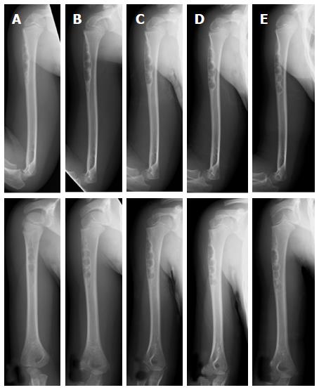
Figure 1 Plain radiographs of a non-ossifying fibroma in the humerus of a 4-year-old female.
An osteolytic lesion is shown at the cortex in the proximal humerus (A). Radiographs taken after 7 mo (B), 1 year (C), 1 year and 7 mo (D), and 2 years and 7 mo (E) together reveal the location of the lesion became more distal with growth of the child. The size of the lesion increased as it slowly ossified (plain radiographs: Anteroposterior view, top; lateral view, bottom).
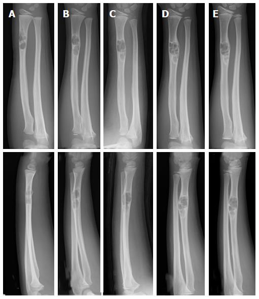
Figure 2 Plain radiographs of a non-ossifying fibroma in the radius of a 9-year-old male.
At the initial assessment an osteolytic lesion in the distal radius is shown with an osteosclerotic rim. Fracture of the irregular adjacent cortex is revealed (A). Radiographs taken after 11 mo (B), 2 years and 1 mo (C), 2 years and 5 mo (D), and 3 years (E). The size of the lesion increased and ossification at the distal end was observed (plain radiographs: Anteroposterior view, top; lateral view, bottom).
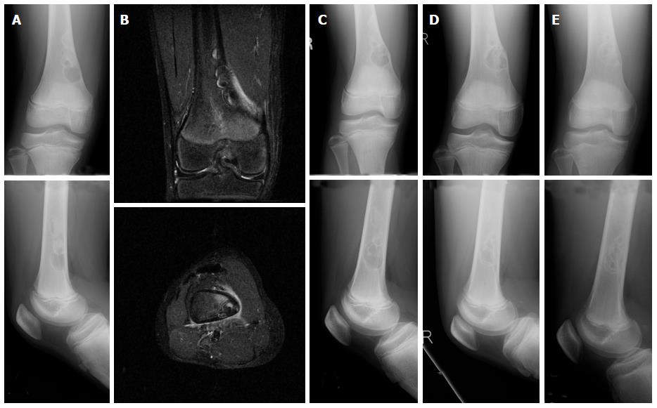
Figure 3 Plain radiograph and MRI of a NOF in the femur of a 13-year-old male who sustained a fracture.
The plain radiographs reveal a multinodular lesion located at the medial posterior part of the distal femur. The lesion is osteolytic at the distal end and ossified at the proximal end. The lesion has expanded at the medial cortex (A). T2-weighted fat-suppression MRIs show high signal intensity and suggest the presence of a fracture (B) (coronal, top; axial, bottom). Radiographs taken after 1 year (C), 1 year and 8 mo (D), and 2 years and 8 mo (E); these radiographs reveal the lesion had enlarged as well as ossified (plain radiographs: anteroposterior view, top; lateral view, bottom) .
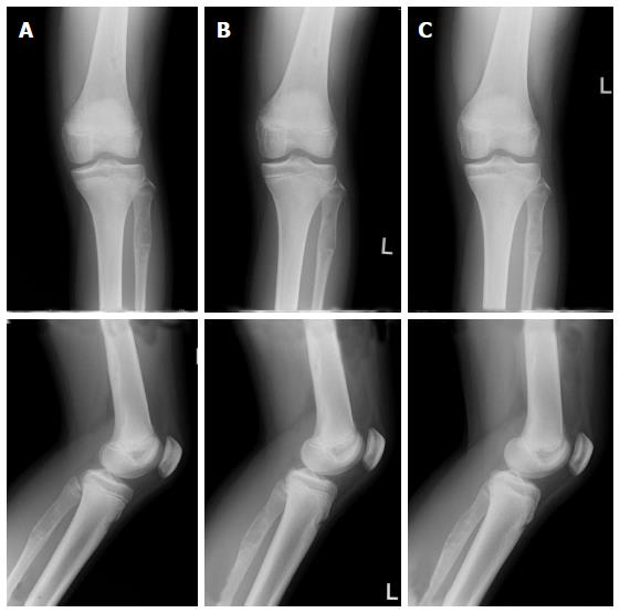
Figure 4 Plain radiographs of non-ossifying fibromas in the femur and the fibula of a 13-year-old female.
The radiographs reveal an osteolytic lesion in the proximal fibula, as well as a NOF in the distal femur (A). Radiographs taken after 9 mo (B) and 1 year and 9 mo (C); the femoral and fibular lesions ossified (plain radiographs: Anteroposterior view, top; lateral view, bottom).
- Citation: Sakamoto A, Arai R, Okamoto T, Matsuda S. Non-ossifying fibromas: Case series, including in uncommon upper extremity sites. World J Orthop 2017; 8(7): 561-566
- URL: https://www.wjgnet.com/2218-5836/full/v8/i7/561.htm
- DOI: https://dx.doi.org/10.5312/wjo.v8.i7.561


