Copyright
©The Author(s) 2017.
World J Orthop. Jul 18, 2017; 8(7): 561-566
Published online Jul 18, 2017. doi: 10.5312/wjo.v8.i7.561
Published online Jul 18, 2017. doi: 10.5312/wjo.v8.i7.561
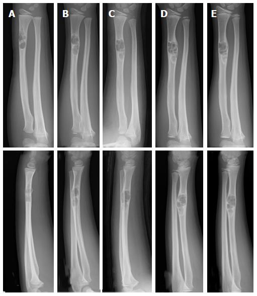
Figure 2 Plain radiographs of a non-ossifying fibroma in the radius of a 9-year-old male.
At the initial assessment an osteolytic lesion in the distal radius is shown with an osteosclerotic rim. Fracture of the irregular adjacent cortex is revealed (A). Radiographs taken after 11 mo (B), 2 years and 1 mo (C), 2 years and 5 mo (D), and 3 years (E). The size of the lesion increased and ossification at the distal end was observed (plain radiographs: Anteroposterior view, top; lateral view, bottom).
- Citation: Sakamoto A, Arai R, Okamoto T, Matsuda S. Non-ossifying fibromas: Case series, including in uncommon upper extremity sites. World J Orthop 2017; 8(7): 561-566
- URL: https://www.wjgnet.com/2218-5836/full/v8/i7/561.htm
- DOI: https://dx.doi.org/10.5312/wjo.v8.i7.561


