Copyright
©The Author(s) 2015.
World J Orthop. Jul 18, 2015; 6(6): 498-504
Published online Jul 18, 2015. doi: 10.5312/wjo.v6.i6.498
Published online Jul 18, 2015. doi: 10.5312/wjo.v6.i6.498
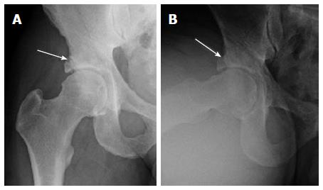
Figure 1 AP (A) and Dunn lateral (B) views of the right hip show right femoral head and neck bump and superolateral acetabular over coverage with suggestion of a rim fracture (arrows).
Notice mild subchondral acetabular sclerosis.
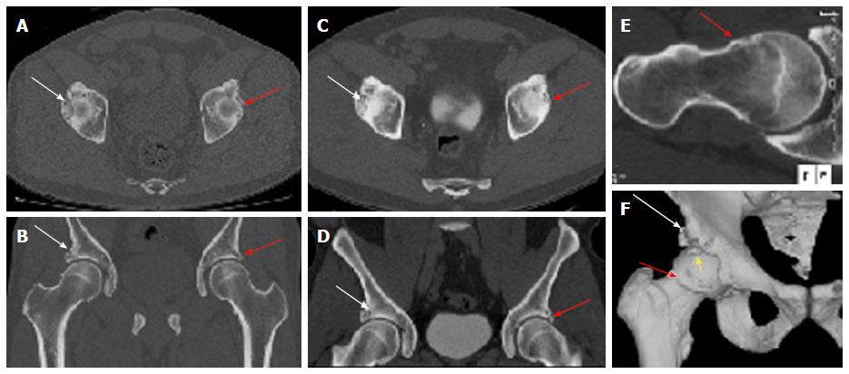
Figure 2 3D computed tomography imaging of the pelvis.
Computed tomography pelvis obtained at current presentation (A, B) and 2 years before (C, D) confirm the unchanged bilateral mixed femoroacetabular impingement anatomy with a chronic right acetabular rim fracture (white arrows) and small left Os acetabulum/labral calcification (yellow arrow). Oblique axial thick slab maximum intensity projection reconstruction (E) along the right femoral neck axis shows the CAM deformity (red arrow) and fibrocystic change at the head and neck junction. Surface rendered 3D bone reconstruction (F) confirms the rim fracture (white arrows) and the CAM deformity (red arrow). Also note loose fragment anteriorly and superiorly, which was subsequently removed on surgery (yellow arrow).
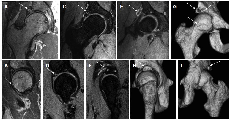
Figure 3 3D magnetic resonance imaging of the right hip.
Multiplanar isotropic reconstructions from 3D fast spin echo proton density weighted (PDW) (A, B) and fat suppressed PDW (C-F) show the acetabular rim fracture (arrows in A, C, F) with pseudoarthrosis and cystic changes; paralabral cyst wrapping around the rectus femoris tendon (arrows in E, F) and CAM deformity (arrow in B). 3D surface rendered bone reconstructions show the bony changes akin to the computed tomography (CT) images with a CAM deformity and bone fragments (arrows in G) and the rim fracture (arrows in H, I), as with 3D CT.
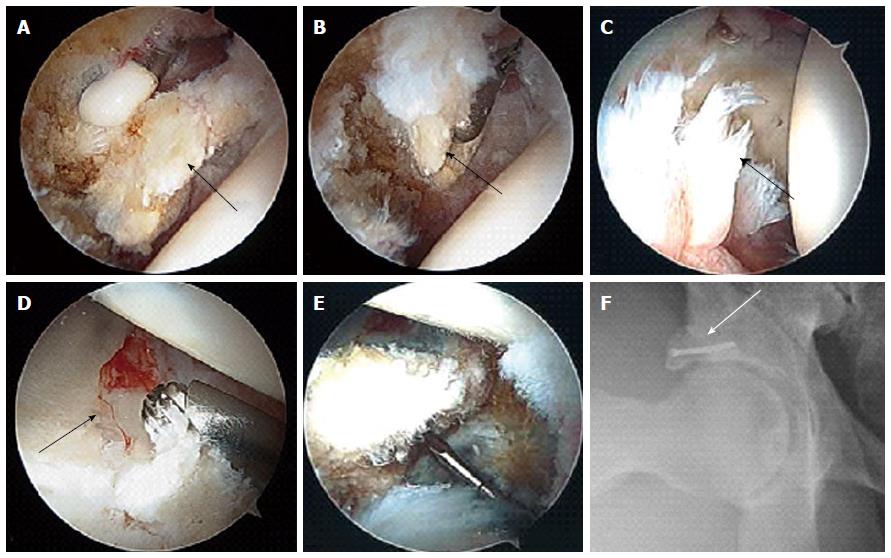
Figure 4 Arthroscopic and follow up images.
Intra-operative photos show the anterior superior acetabular loose fragment (arrow in A) being freed with a radiofrequency device and then removed with an arthroscopic grasper via the mid-anterior portal (arrow in B). Notice the shredded labrum that remained anteriorly (arrow in C). A 4.5 mm burr pictured above the lateral rim fracture fragment. The crack in articular cartilage can be seen running from anterior to posterior (arrow in D). This rim fragment was fixed arthroscopically using a 2.4 mm headless screw (E). Follow up Dunn view shows the nicely fixed acetabular rim fracture with the screw (arrow in F).
- Citation: Chhabra A, Nordeck S, Wadhwa V, Madhavapeddi S, Robertson WJ. Femoroacetabular impingement with chronic acetabular rim fracture - 3D computed tomography, 3D magnetic resonance imaging and arthroscopic correlation. World J Orthop 2015; 6(6): 498-504
- URL: https://www.wjgnet.com/2218-5836/full/v6/i6/498.htm
- DOI: https://dx.doi.org/10.5312/wjo.v6.i6.498


