Copyright
©The Author(s) 2015.
World J Orthop. Jul 18, 2015; 6(6): 498-504
Published online Jul 18, 2015. doi: 10.5312/wjo.v6.i6.498
Published online Jul 18, 2015. doi: 10.5312/wjo.v6.i6.498
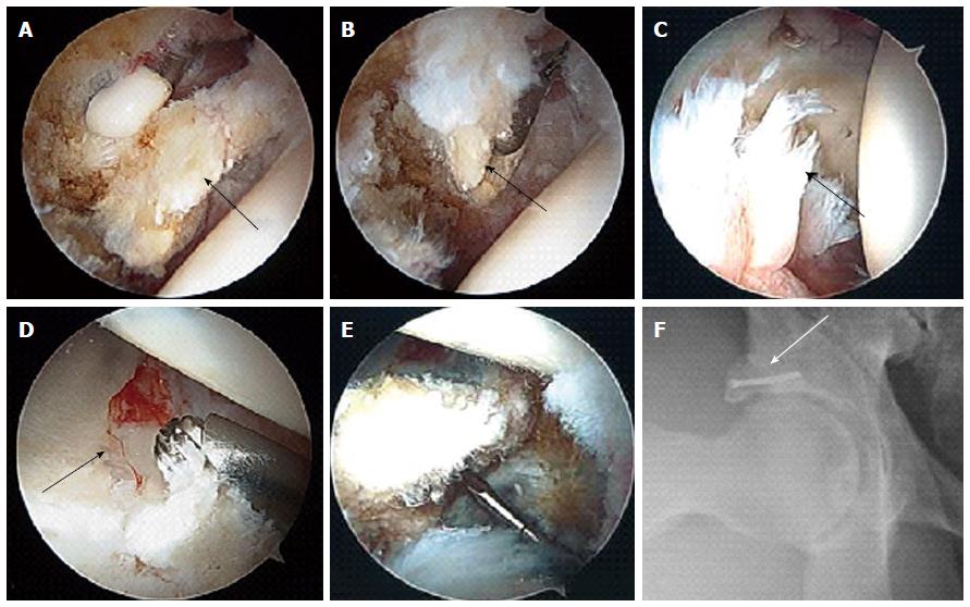
Figure 4 Arthroscopic and follow up images.
Intra-operative photos show the anterior superior acetabular loose fragment (arrow in A) being freed with a radiofrequency device and then removed with an arthroscopic grasper via the mid-anterior portal (arrow in B). Notice the shredded labrum that remained anteriorly (arrow in C). A 4.5 mm burr pictured above the lateral rim fracture fragment. The crack in articular cartilage can be seen running from anterior to posterior (arrow in D). This rim fragment was fixed arthroscopically using a 2.4 mm headless screw (E). Follow up Dunn view shows the nicely fixed acetabular rim fracture with the screw (arrow in F).
- Citation: Chhabra A, Nordeck S, Wadhwa V, Madhavapeddi S, Robertson WJ. Femoroacetabular impingement with chronic acetabular rim fracture - 3D computed tomography, 3D magnetic resonance imaging and arthroscopic correlation. World J Orthop 2015; 6(6): 498-504
- URL: https://www.wjgnet.com/2218-5836/full/v6/i6/498.htm
- DOI: https://dx.doi.org/10.5312/wjo.v6.i6.498


