Copyright
©The Author(s) 2015.
World J Orthop. Jul 18, 2015; 6(6): 498-504
Published online Jul 18, 2015. doi: 10.5312/wjo.v6.i6.498
Published online Jul 18, 2015. doi: 10.5312/wjo.v6.i6.498
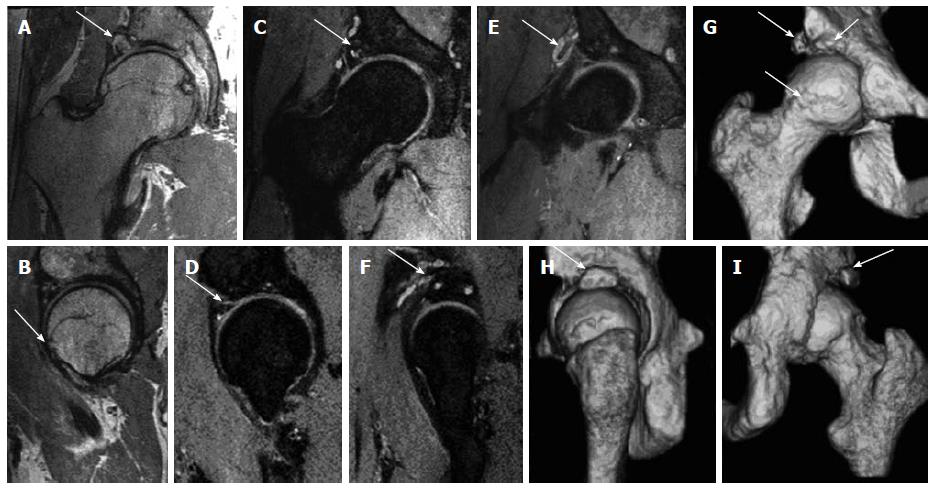
Figure 3 3D magnetic resonance imaging of the right hip.
Multiplanar isotropic reconstructions from 3D fast spin echo proton density weighted (PDW) (A, B) and fat suppressed PDW (C-F) show the acetabular rim fracture (arrows in A, C, F) with pseudoarthrosis and cystic changes; paralabral cyst wrapping around the rectus femoris tendon (arrows in E, F) and CAM deformity (arrow in B). 3D surface rendered bone reconstructions show the bony changes akin to the computed tomography (CT) images with a CAM deformity and bone fragments (arrows in G) and the rim fracture (arrows in H, I), as with 3D CT.
- Citation: Chhabra A, Nordeck S, Wadhwa V, Madhavapeddi S, Robertson WJ. Femoroacetabular impingement with chronic acetabular rim fracture - 3D computed tomography, 3D magnetic resonance imaging and arthroscopic correlation. World J Orthop 2015; 6(6): 498-504
- URL: https://www.wjgnet.com/2218-5836/full/v6/i6/498.htm
- DOI: https://dx.doi.org/10.5312/wjo.v6.i6.498


