Copyright
©2014 Baishideng Publishing Group Inc.
World J Orthop. Nov 18, 2014; 5(5): 653-659
Published online Nov 18, 2014. doi: 10.5312/wjo.v5.i5.653
Published online Nov 18, 2014. doi: 10.5312/wjo.v5.i5.653
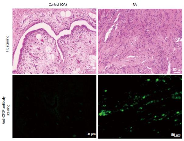
Figure 1 Connective tissue growth factor expression was increased at synovial tissue in rheumatoid arthritis.
Representative results of hematoxylin and eosin (HE) staining, immunofluorescence anti-connective tissue growth factor (CTGF) antibody staining, and anti-F4/80 antibody staining are shown using surgical samples from patients with rheumatoid arthritis (RA) and osteoarthritis (OA). The observed CTGF expression was stronger in the samples of patients with RA than in the samples of patients with OA.
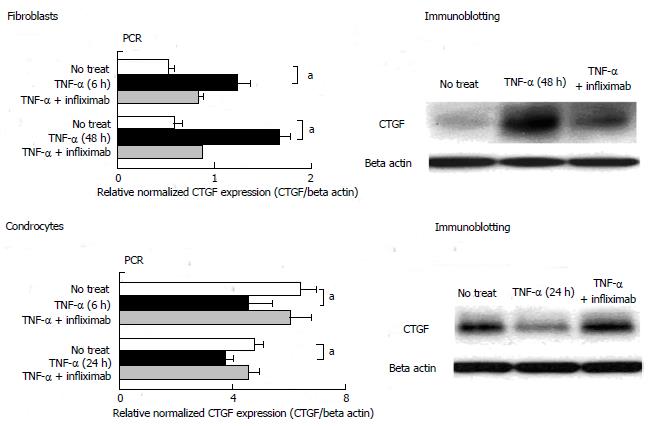
Figure 2 Tumor necrosis factor-α positively regulated connective tissue growth factor production in synovial fibroblasts and negatively regulated connective tissue growth factor production in chondrocytes.
Connective tissue growth factor (CTGF) production from the human synovial fibroblasts cell line (MH7A) and the human chondrocytes cell line (OUMS-27) stimulated with/without tumor necrosis factor (TNF)-α were evaluated by immunoblotting and quantitative real time polymerase chain reaction (PCR). TNF-α promoted CTGF production by synovial fibroblasts and inhibited the production by chondrocytes. Statistical analysis (paired t test) was performed, and aP < 0.05 were considered to be statistically significant. aP < 0.05, TNF-αvs no treat.
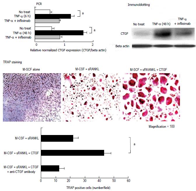
Figure 3 Connective tissue growth factor enhanced macrophage-colony stimulating factor/ receptor activator of nuclear factor kappa-B ligand mediated osteoclastgenesis.
Figure 3 Shows images of tartrate-resistant acidic phosphatase (TRAP) staining and the number of TRAP positive cells. For the evaluation of osteoclastogenesis, CD14+ were purified from peripheral blood mononuclear cells of healthy volunteers to obtain osteoclastic progenitor cells. Osteoclasts were induced with macrophage-colony stimulating factor (M-CSF) and soluble receptor activator of nuclear factor kappa-B ligand (sRANKL) and the osteoclastgenesis was evaluated by TRAP staining. The TRAP positive cells were defined as osteoclasts. Connective tissue growth factor (CTGF) alone could not help in the differentiation of osteoclasts (data not shown). M-CSF/RANKL-mediated osteoclastogenesis was enhanced, which was detected by the production of CTGF by larger and higher number of osteoclasts; this enhancing effect was abolished by anti-CTGF antibody. The bars in Figure 2 indicate the standard deviation. Statistical analysis (paired t test) was performed, and P values < 0.05 were considered to be statistically significant. aP < 0.05, M-CSF + sRANKL + CTGF vs M-CSF + sRANKL.
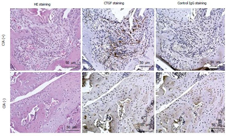
Figure 4 Increased in vivo expression of connective tissue growth factor at the articular tissue in collagen-induced arthritis mice.
The collagen-induced arthritis (CIA) mice were sacrificed at 8 wk after immunization for immunohistochemical analysis. The immunohistochemical staining showed massive connective tissue growth factor (CTGF) expression in the articular tissue samples from CIA mice (indicated by arrow) using anti-CTGF antibody or control goat immunoglobulin G antibody. Serial sections of the articular tissue samples were also counterstained with hematoxylin/eosin (HE) for detection of the arthritis.
Figure 5 Blocking connective tissue growth factor prevented the development of arthritis in collagen-induced arthritis mice.
Mice with collagen-induced arthritis (CIA) were randomly selected and were intraperitoneally administered every week with anti-connective tissue growth factor (CTGF) monoclonal antibodies (mAbs) (white triangle; CIA-anti-CTGF Ab+) or control purified immunoglobulin (white square; CIA-anti-CTGF Ab−) from 1 wk before immunization to 6 weeks after immunization. Each group comprised 12 mice. The mice were monitored for arthritis every week and scored in a blinded manner (A). Blocking CTGF could efficiently prevent the development of CIA in mice. Bars in Figure 3 indicate the standard deviation. Statistical analysis (anti-CTGF Ab+vs anti-CTGF Ab−) was performed, and P values < 0.05 were considered to be statistically significant. aP < 0.05, CIA anti-CTGF Ab vs Control. For evaluation of osteoclastogenesis, CD14+ osteoclastic progenitor cells were purified from splenocytes at 8 wk after immunization and osteoclasts were then induced with macrophage-colony stimulating factor (M-CSF) and soluble receptor activator of nuclear factor kappa-B ligand (sRANKL). Osteoclastogenesis was suppressed in mice with CIA treated using anti-CTGF mAb (B) compared to the non-treated mice. The bars in Figure 3 indicate the standard deviation. Statistical analysis was performed, and P values < 0.05 were considered to be statistically significant. aP < 0.05, CIA anti-CTGF Ab (-) vs Control; CIA anti-CTGF Ab (-) vs CIA anti-CTGF Ab (+).
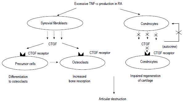
Figure 6 Hypothesis of the role of connective tissue growth factor in the pathogenesis of rheumatoid arthritis.
Hypothesis of the possible role of connective tissue growth factor (CTGF) in the pathogenesis of rheumatoid arthritis (RA). TNF: Tumor necrosis factor.
- Citation: Nozawa K, Fujishiro M, Takasaki Y, Sekigawa I. Inhibition of rheumatoid arthritis by blocking connective tissue growth factor. World J Orthop 2014; 5(5): 653-659
- URL: https://www.wjgnet.com/2218-5836/full/v5/i5/653.htm
- DOI: https://dx.doi.org/10.5312/wjo.v5.i5.653


