Copyright
©2012 Baishideng Publishing Group Co.
World J Orthop. Jul 18, 2012; 3(7): 109-113
Published online Jul 18, 2012. doi: 10.5312/wjo.v3.i7.109
Published online Jul 18, 2012. doi: 10.5312/wjo.v3.i7.109
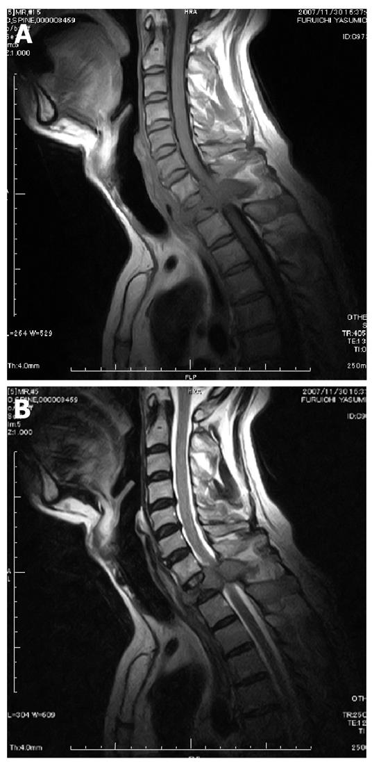
Figure 1 Magnetic resonance imaging revealing compression of the spinal cord at the level of the first thoracic vertebra by a lesion that was considered a metastasis of an unknown primary tumor.
A: T1-weighted image; B: T2-weighted image.
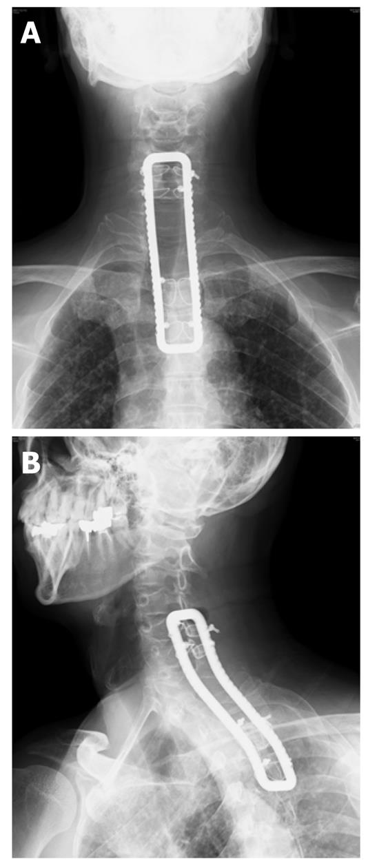
Figure 2 Radiography after first surgery.
The laminectomy of C7, Th1 and Th2 and fixation of C5-Th4 were performed by using a sublaminar wire and a rectangle rod. A: Anteroposterior view; B: Oblique view.
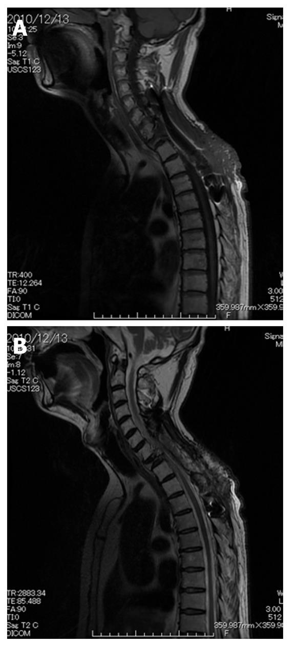
Figure 3 Magnetic resonance imaging 2 years after first surgery.
The pseudomeningocele gradually grew to about 15 cm in diameter from C5 to Th6 level. A: T1-weighted sagittal image; B: T2-weighted sagittal image; C: T2-weighted axial image.
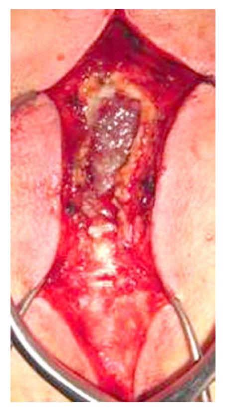
Figure 4 Intraoperative photo before capsule incision of pseudomeningocele.
About 50 mL of colorless clear fluid was released after the capsule incision and no damage to the dura mater was noted.
Figure 5 Magnetic resonance imaging showing the pseudomeningocele had disappeared 3 mo after closure of the cerebrospinal fluid leak.
A: T1-weighted image; B: T2-weighted image.
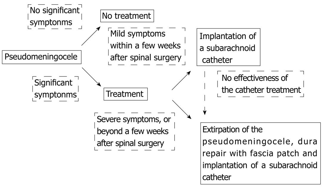
Figure 6 Algorithm for treatment of pseudomeningcele.
Conservative treatment is generally recommended in patients without significant symptoms and surgical treatments should be performed in those with significant symptoms.
- Citation: Srilomsak P, Okuno K, Sakakibara T, Wang Z, Kasai Y. Giant pseudomeningocele after spinal surgery: A case report. World J Orthop 2012; 3(7): 109-113
- URL: https://www.wjgnet.com/2218-5836/full/v3/i7/109.htm
- DOI: https://dx.doi.org/10.5312/wjo.v3.i7.109


