Copyright
©2011 Baishideng Publishing Group Co.
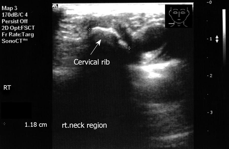
Figure 1 Transverse imaging of the right neck region.
Note the echogenic structure with acoustic shadow.
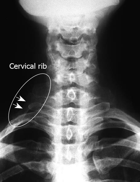
Figure 2 X-ray corresponding to Figure 1.
Note the cervical rib.
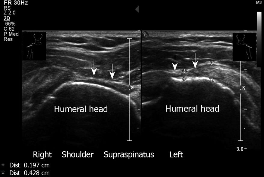
Figure 3 Partial thickness tear of supraspinatus.
Right and left shoulder. Note the narrowing of the tendon. Transverse sonogram. A 70-year old male who presents with bilateral shoulder pain and has a painful arc at clinical examination.
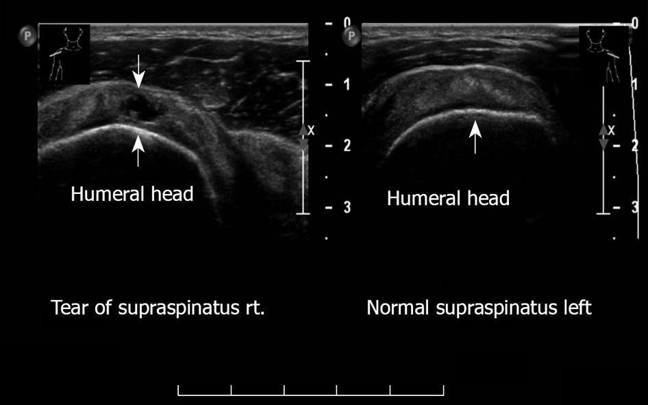
Figure 4 Transverse sonogram, supraspinatus right and left shoulder.
Anechoic defect. Partial thickness tear, right. Left: Normal supraspinatus.
Figure 5 Massive full-thickness tear of supraspinatus.
Left shoulder. Non visualization of tendon. This 81-year old female has severe shoulder pain that increases at night.
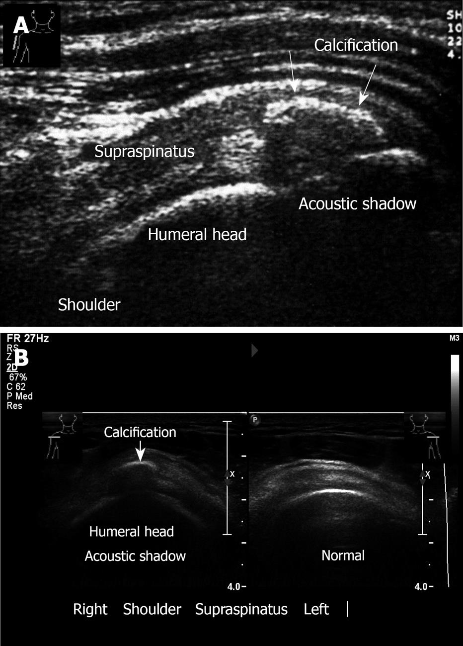
Figure 6 Shoulder calcification.
A: Calcific tendinitis. Calcification in the supraspinatus tendon, longitudinal view. Note: Acoustic shadow behind the calcification. A 29-year old woman presents with a short history (3 d) of incapacitating shoulder pain and with severe restriction in the range of shoulder movement; B: Transverse sonogram. Calcification of supraspinatus, right. Left normal sonogram.
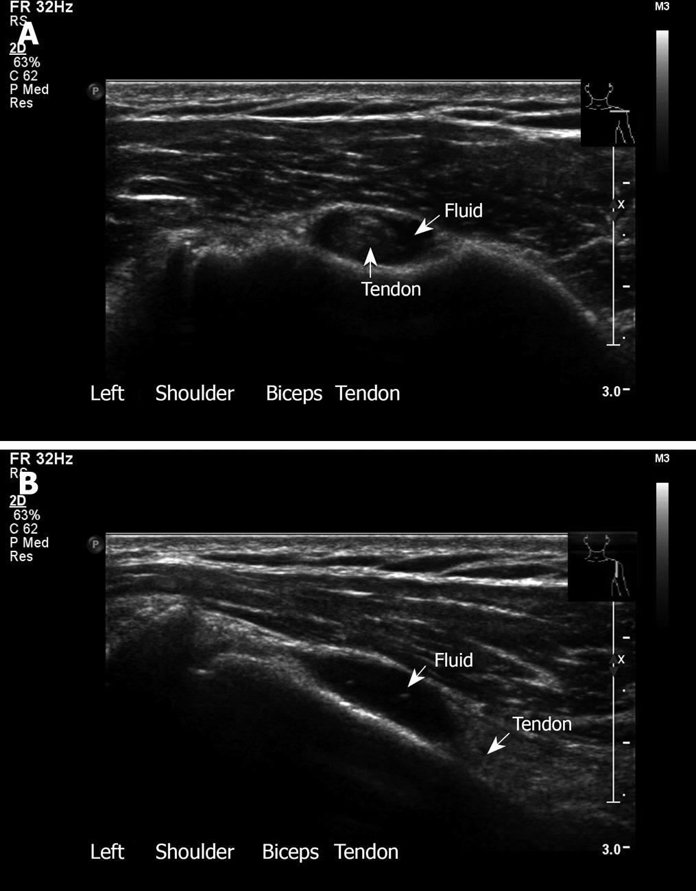
Figure 7 Biceps tendon.
A: Bicipital tendinitis. Transverse sonogram. The tendon is surrounded by fluid. This 33-year old woman presents with pain and local tenderness in the area of the bicipital groove; B: Longitudinal view with fluid around the biceps.
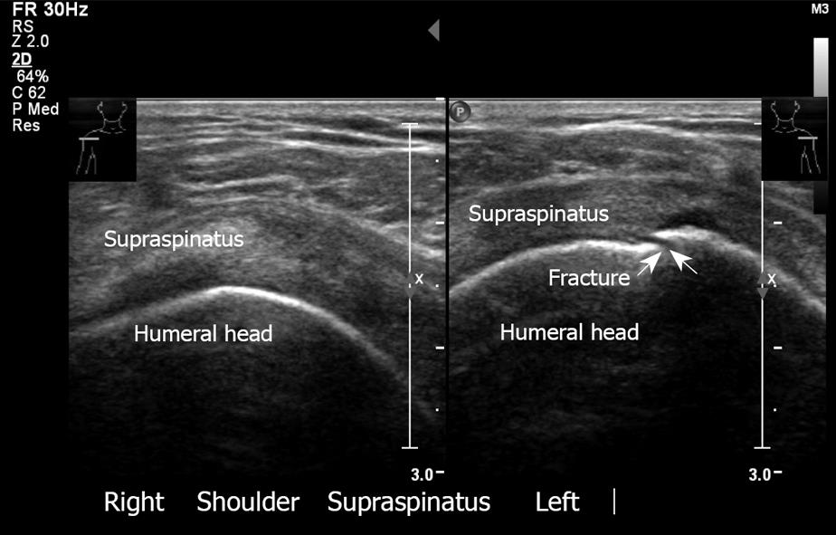
Figure 8 Fracture of greater tuberosity, left.
Note the discontinuity of the bone. The right shoulder has normal appearance. Transverse sonogram.
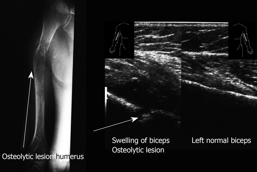
Figure 9 Longitudinal sonogram.
Swelling of soft tissue with severe irregularity of the cortex right humerus. Left biceps normal. X-ray of the right arm demonstrates the osteolytic lesion of the right humerus. A 75-year old man presents with swelling of the right humerus, limitation of movement, night pain and weight loss.
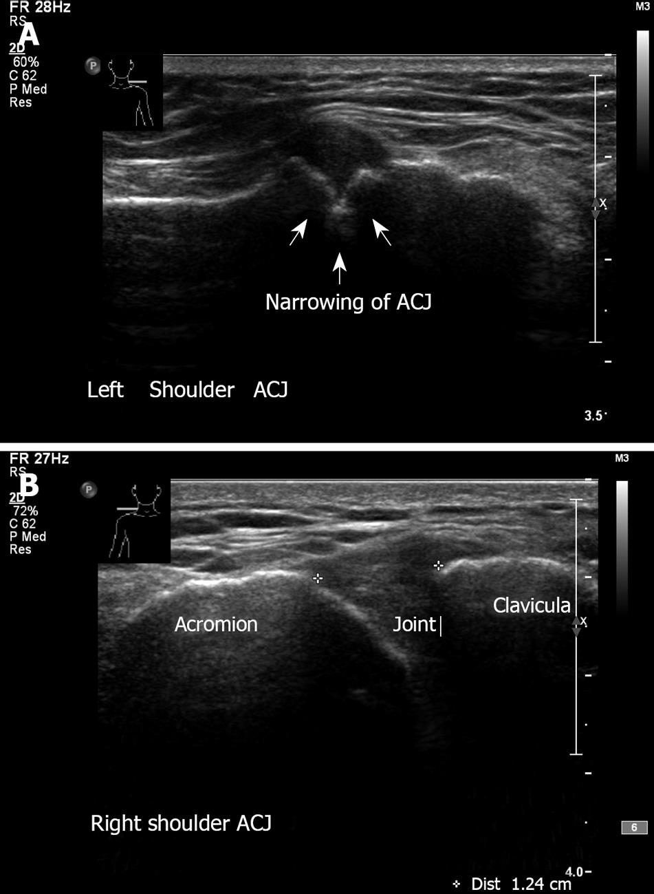
Figure 10 Acromioclavicular joint.
A: Left shoulder - A-C arthritis. Note the irregularity and narrowing of the acromioclavicular joint. Erosive changes at the articular surface of the joint. A 65-year old man with a long history of pain and tenderness related to the acromioclavicular joint; B: Normal right acromioclavicular joint with open joint space.
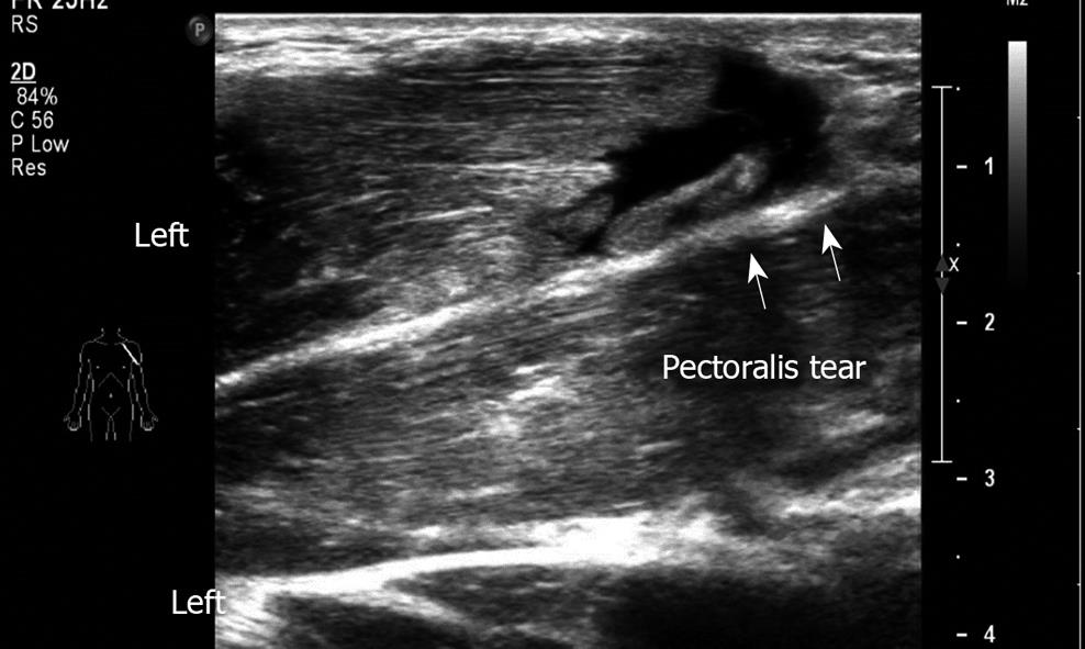
Figure 11 Longitudinal sonogram.
Demonstrates a large pectoralis tear left. A 23-year old soldier felt a pop when he was lifting a wounded friend.
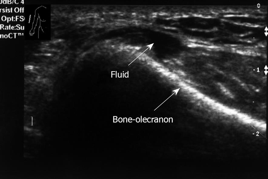
Figure 12 Longitudinal sonogram, right elbow.
Small amount of fluid posterior aspect. No erosive changes in the bone.
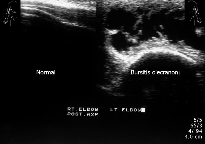
Figure 13 Longitudinal view, left elbow.
Swelling and fluid of soft tissue. Reactive elbow joint effusion, corresponding to bursitis. Right elbow normal appearance. This 60-year old man had pain and swelling over the dorsal aspect of the left elbow, olecranon bursitis.
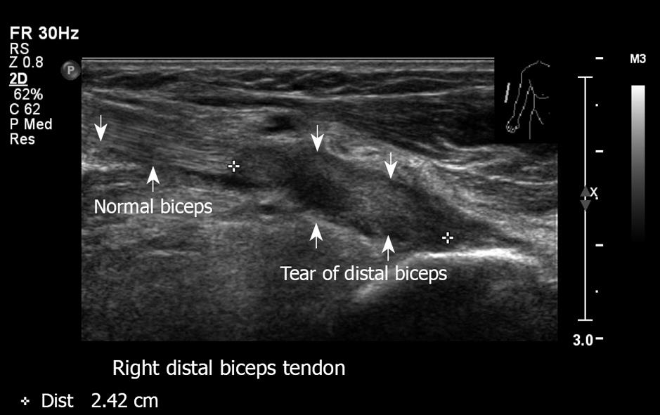
Figure 14 Right distal biceps tendon tear in the area of insertion.
Proximal biceps look normal. This 62-year old man felt a severe sudden sharp pain in the distal humerus region after lifting a heavy suitcase.
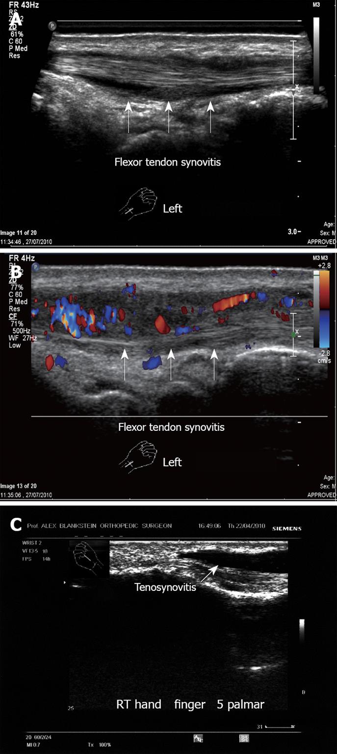
Figure 15 Tenosynovitis.
A: Left hand flexor tendon synovitis. Note the fluid around the tendon. No tear is demonstrated; B: Left hand flexor tendon synovitis. Note the hyper-vascularity with the vascular inflammation signs; C: Flexor tendon synovitis, right hand mid-phalanx. Note the large amount of clear fluid around the tendon. This 55-year old woman presented with acute pain in the palmar aspect of the hand with irregular synovial thickening, increased fluid and hypervascularity.
Figure 16 Anterior knee.
A: Left knee longitudinal view. Transducer on the anterior aspect of the knee. Note the fluid in the suprapatellar recess. Normal quadriceps tendon with its insertion to the patella; B: Similar transducer position demonstrating synovial hypertrophy. A 68-year old woman with rheumatoid arthritis presented with pain and swelling of the right anterior knee.
Figure 17 Left knee medial aspect.
Ultrasound demonstrates medial collateral ligament. Joint space is normal. Neutral position. Joint space measures 0.656 cm. The same area under valgus stress, demonstrating joint space of 0.947 cm, pointing to medial collateral ligament insufficiency, clinically manifested by mild knee instability.
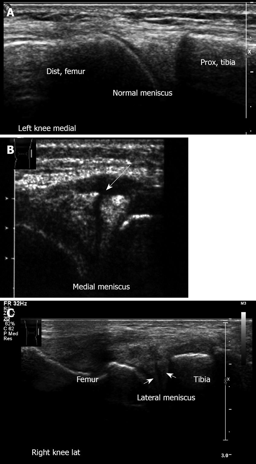
Figure 18 Meniscal lesions.
A: Left knee medial aspect, longitudinal sonogram. Medial meniscus anterior horn. Note the triangle-shaped hyperechoic structure of the normal medial meniscus; B: Medial meniscus lesion. Note the cleft and irregularity of the torn meniscus. This young football player suffered an acute twisting injury. Clinically he had pain with mild swelling on the medial aspect, there was a joint effusion with medial line tenderness. Mac-Murray and Apley tests were positive over the medial meniscus; Right knee lateral meniscus lesion. Note the irregularity of the torn lateral meniscus.
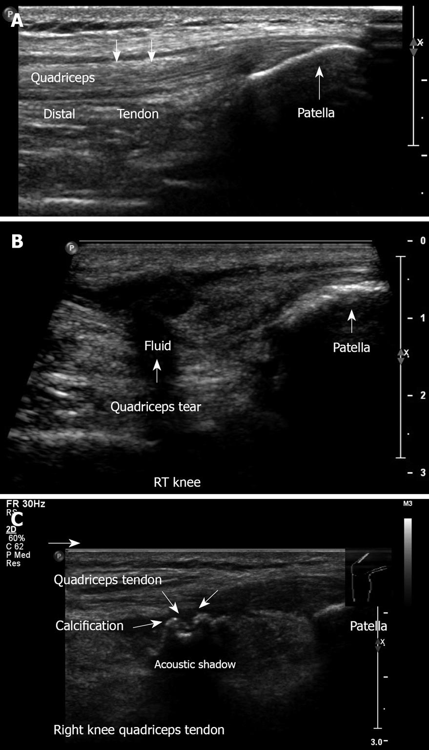
Figure 19 Anterior knee.
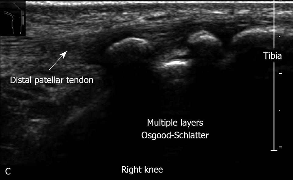
A: Longitudinal ultrasound image obtained in the midline, demonstrating the anterior knee, quadriceps tendon with its insertion to the patella, suprapatellar recess, and the patella. No effusion is visible; B: Longitudinal view. Complete quadriceps tendon tear. This 62-year old physician suffered direct trauma to his right knee when falling down stairs; C: Quadriceps tendinitis with calcification of the right knee. This 50-year old man had a contusion with hematoma of the quadriceps muscle one year ago.
Figure 20 Right knee Osgood-Schlatter disease.
Note the severe irregularity of tibial tuberosity. This 14-year old football player has severe pain and swelling of the tibial tuberosity,
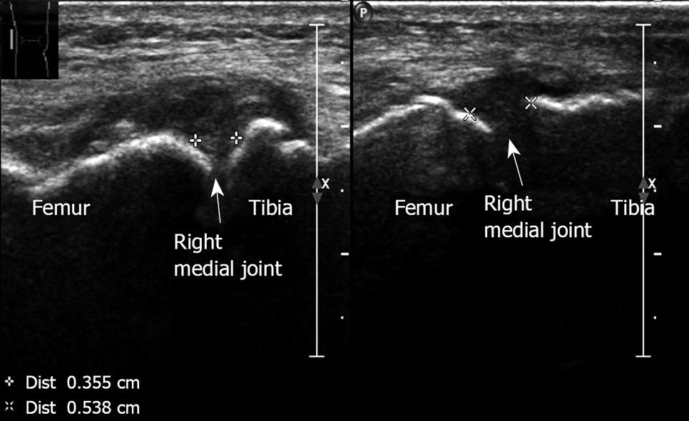
Figure 21 Osteoarthritis medial aspect, right knee.
Medial joint space narrowing with osteophyte formation and thickening of the medial collateral ligament, measured 0.355 cm. Lateral joint with normal appearance measured 0.538 cm. A 75-year old woman with typical findings of osteoarthritic changes.
Figure 22 Longitudinal sonogram of the thigh.
A young basketball player presented with pain over the anterior aspect of the distal right femur. Note the irregularity with partial tear of the muscle. Left thigh: Normal appearance.
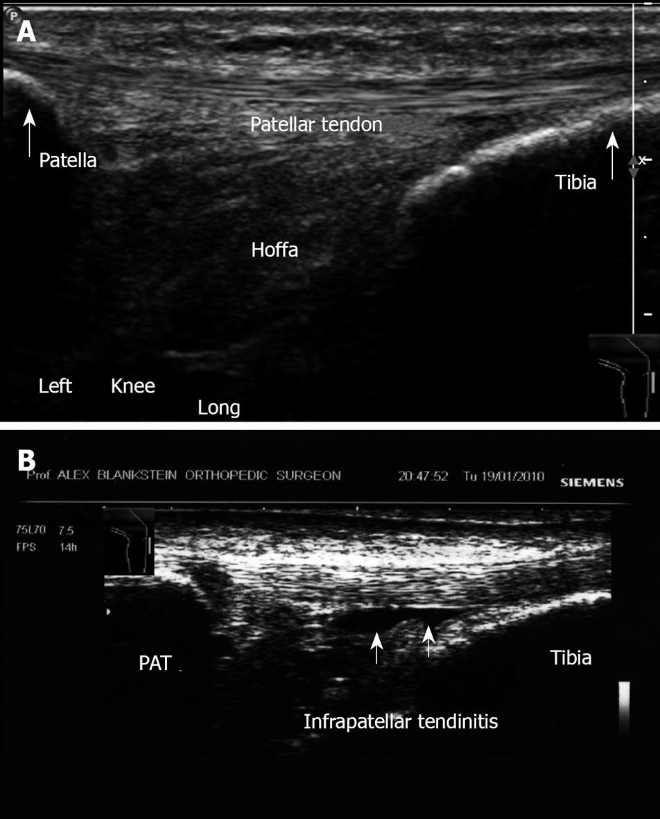
Figure 23 Patellar tendon.
A: Longitudinal view. Anterior aspect. Ultrasound image shows the patellar tendon from its origin in the patella into the tibial tuberosity left knee; B: Infrapatellar tendinitis. Note fluid accumulation deep to the patellar tendon.
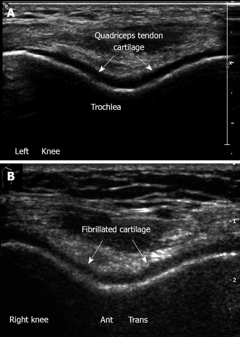
Figure 24 Femoral throchlea.
A: Left knee. Flexion position. Anterior transverse view. Note trochlear cartilage of femur. The hyaline cartilage is a hypoechoic homogenous structure with sharp margins, overlying the bright hyperechoic line of subchondral bone; B: Cartilage lesion. Anterior transverse right knee in flexion, irregularity and narrowing of the hyaline cartilage which is roughened and fibrillated.
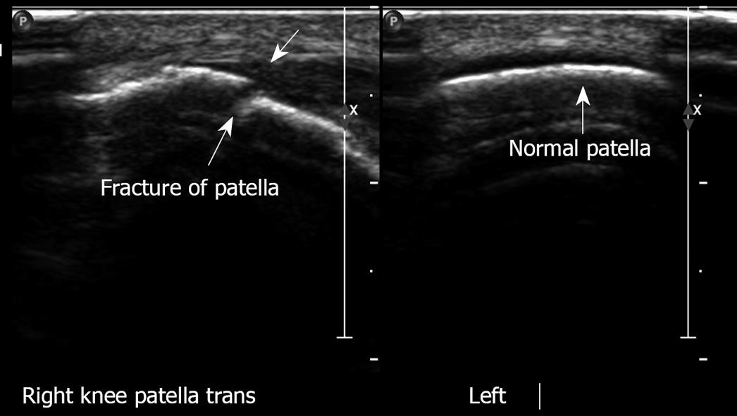
Figure 25 Longitudinal view, right patella with fracture.
Left patella normal.
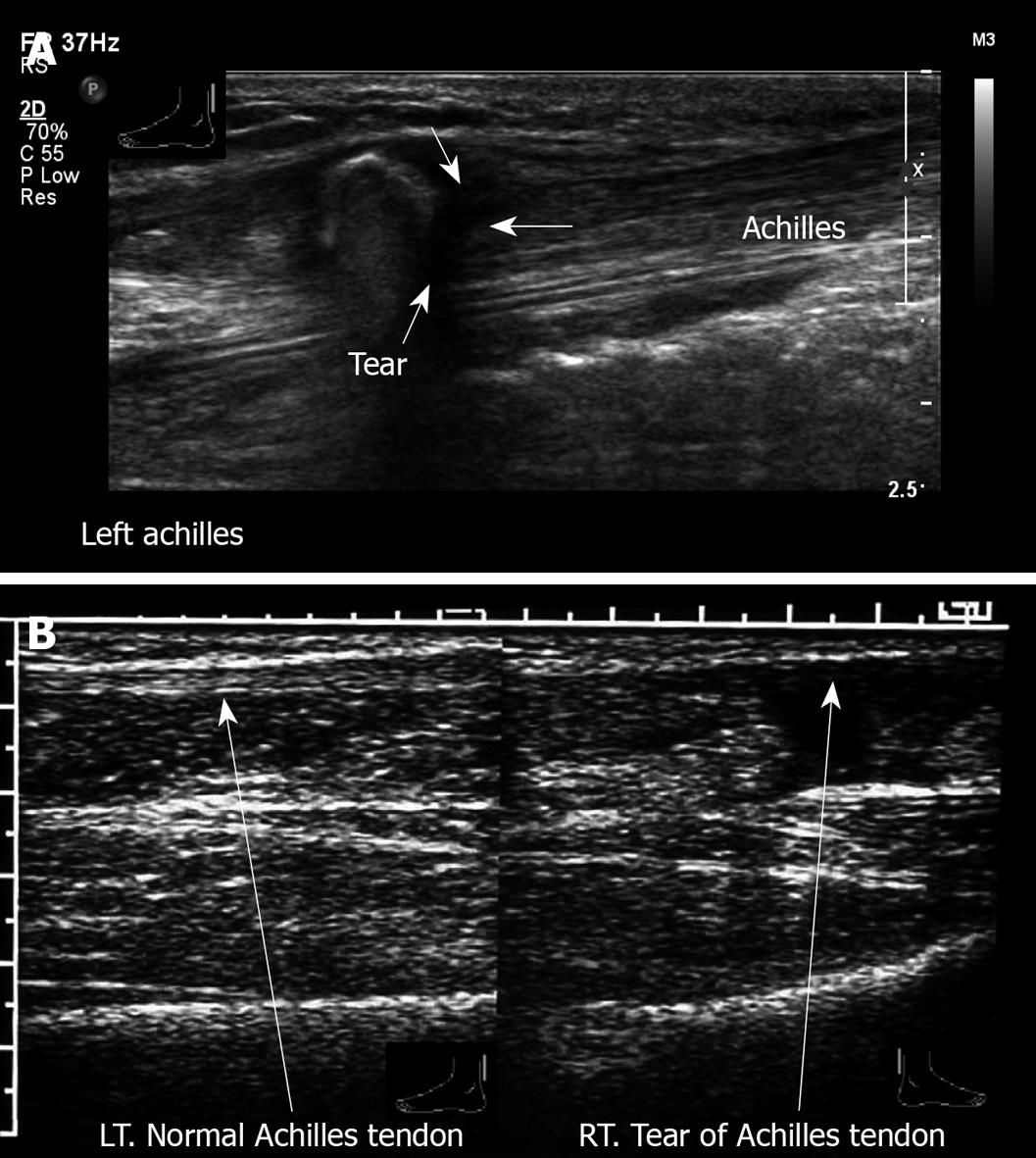
Figure 26 Achilles tendon.
A: Longitudinal view, left ankle posterior aspect. Complete tear of Achilles tendon with retraction. A 54-year old man felt a sudden sharp pain in the left Achilles tendon while running. Physical examination revealed absence of plantar flexion and a positive Thompson test; B: Longitudinal view. Right: tear of Achilles tendon. Left: Normal Achilles tendon.
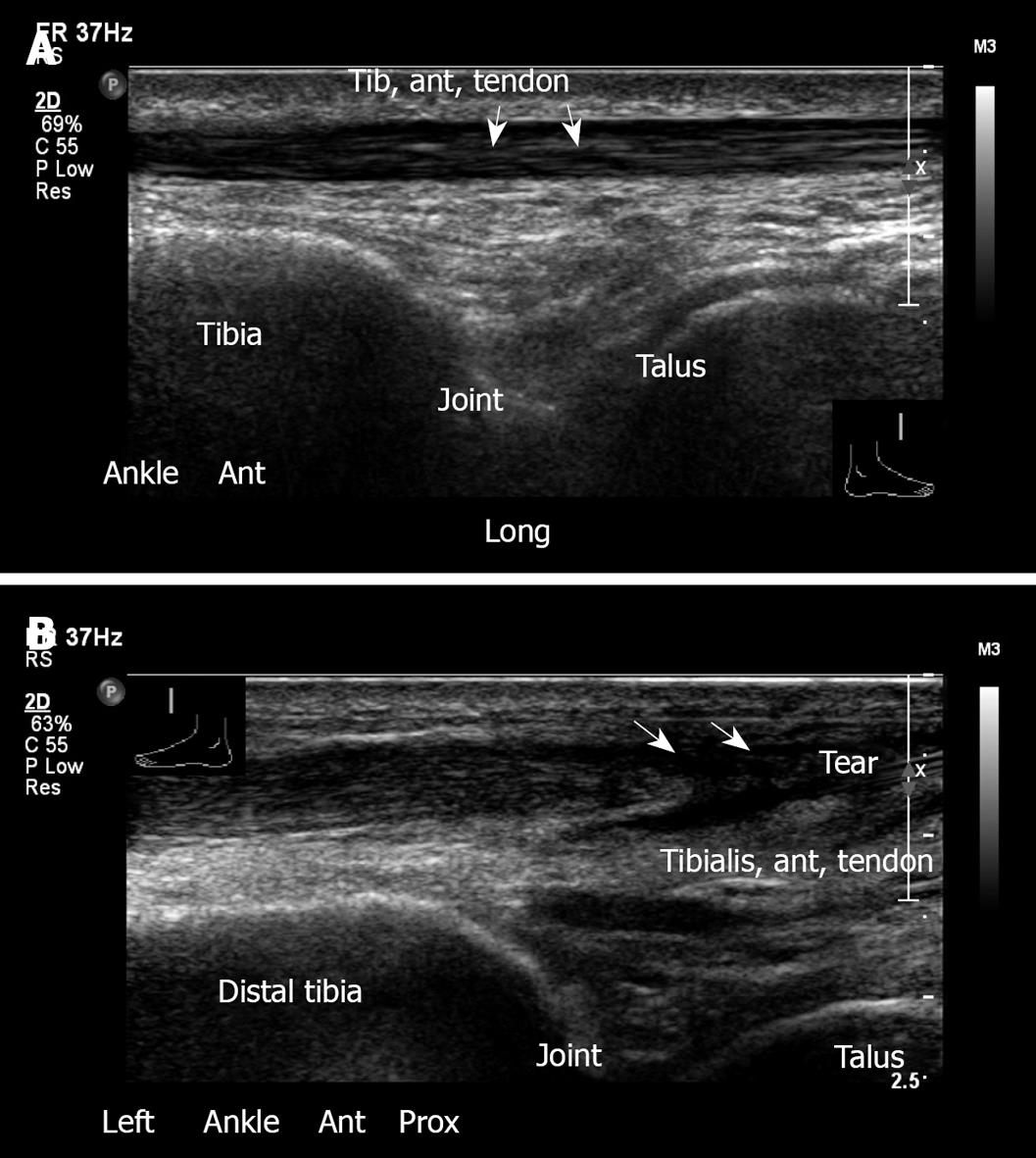
Figure 27 Ankle joint.
A: Anterior longitudinal view, ankle. Tibiotalar joint with normal appearance of tibialis anterior tendon; B: Left ankle, longitudinal view. Tear of tibialis anterior tendon. This 68-year old male suffered from pain in the anterior aspect of the left ankle after much walking. No trauma had occurred.
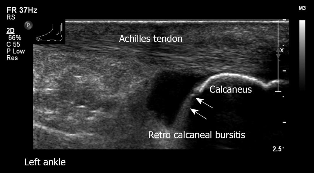
Figure 28 Left ankle retro-calcaneal bursitis.
Longitudinal view. Normal Achilles tendon. Note the large amount of fluid in the retro-calcaneal bursa.
Figure 29 Longitudinal view, distal right leg.
Swelling of soft tissue with fluid. Note increased echogenicity and thickening of the subcutaneous fat in the inflamed region.
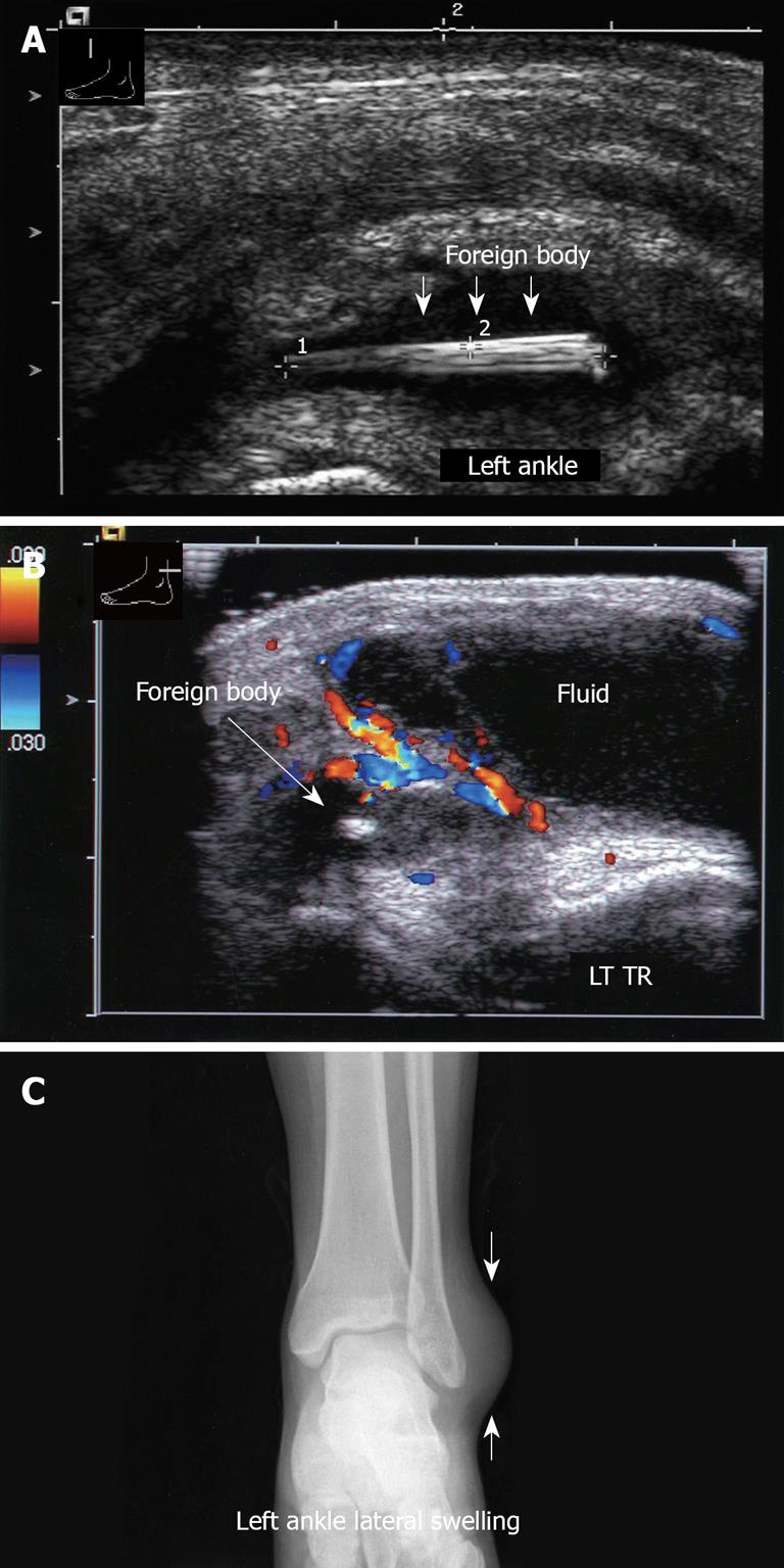
Figure 30 Ankle joint X-ray and US images.
A: Longitudinal sonogram, left ankle, demonstrates a wooden foreign body; B: Transverse view, left ankle. Note the hypervascularity in the inflamed area; C: Corresponding X-ray of left ankle. Note the swelling on the lateral aspect. No foreign body is visible.
- Citation: Blankstein A. Ultrasound in the diagnosis of clinical orthopedics: The orthopedic stethoscope. World J Orthop 2011; 2(2): 13-24
- URL: https://www.wjgnet.com/2218-5836/full/v2/i2/13.htm
- DOI: https://dx.doi.org/10.5312/wjo.v2.i2.13


