Copyright
©2011 Baishideng Publishing Group Co.
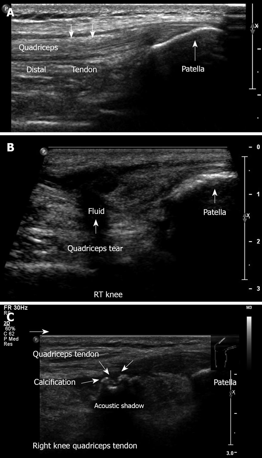
Figure 19 Anterior knee.
A: Longitudinal ultrasound image obtained in the midline, demonstrating the anterior knee, quadriceps tendon with its insertion to the patella, suprapatellar recess, and the patella. No effusion is visible; B: Longitudinal view. Complete quadriceps tendon tear. This 62-year old physician suffered direct trauma to his right knee when falling down stairs; C: Quadriceps tendinitis with calcification of the right knee. This 50-year old man had a contusion with hematoma of the quadriceps muscle one year ago.
- Citation: Blankstein A. Ultrasound in the diagnosis of clinical orthopedics: The orthopedic stethoscope. World J Orthop 2011; 2(2): 13-24
- URL: https://www.wjgnet.com/2218-5836/full/v2/i2/13.htm
- DOI: https://dx.doi.org/10.5312/wjo.v2.i2.13


