Copyright
©2011 Baishideng Publishing Group Co.
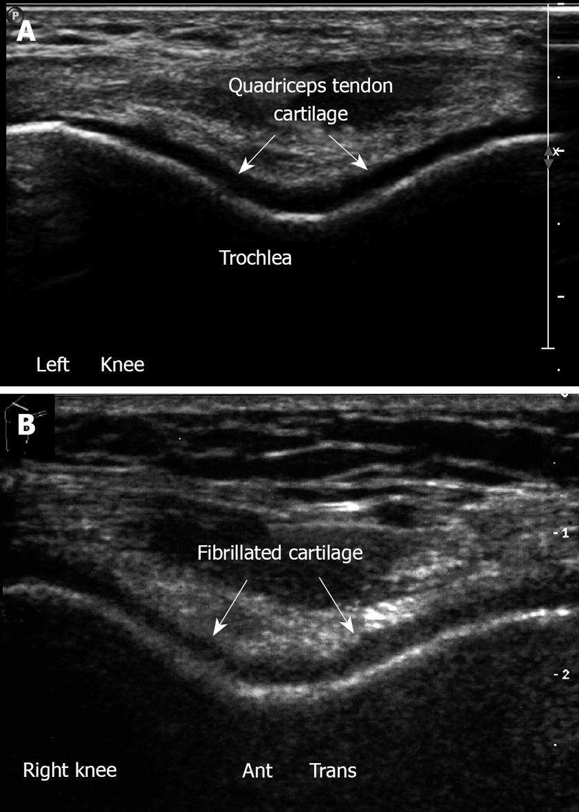
Figure 24 Femoral throchlea.
A: Left knee. Flexion position. Anterior transverse view. Note trochlear cartilage of femur. The hyaline cartilage is a hypoechoic homogenous structure with sharp margins, overlying the bright hyperechoic line of subchondral bone; B: Cartilage lesion. Anterior transverse right knee in flexion, irregularity and narrowing of the hyaline cartilage which is roughened and fibrillated.
- Citation: Blankstein A. Ultrasound in the diagnosis of clinical orthopedics: The orthopedic stethoscope. World J Orthop 2011; 2(2): 13-24
- URL: https://www.wjgnet.com/2218-5836/full/v2/i2/13.htm
- DOI: https://dx.doi.org/10.5312/wjo.v2.i2.13


