Copyright
©The Author(s) 2021.
World J Orthop. May 18, 2021; 12(5): 254-269
Published online May 18, 2021. doi: 10.5312/wjo.v12.i5.254
Published online May 18, 2021. doi: 10.5312/wjo.v12.i5.254
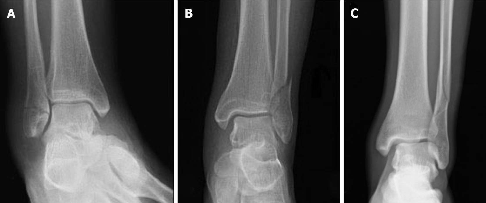
Figure 1 Danis-Weber classification of distal fibula isolated fractures.
A: Type A; B: Type B; C: Type C.
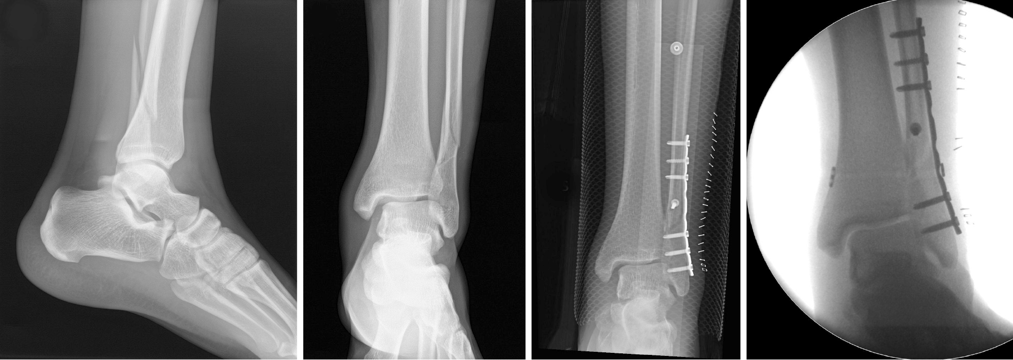
Figure 2 Lateral, mortise and antero-posterior radiographic views of a Danis-Weber type B left distal fibula fracture.
Conservative treatment is the correct choice for this case because minimal displacement of the fracture and absence of associated ankle instability are demonstrated.
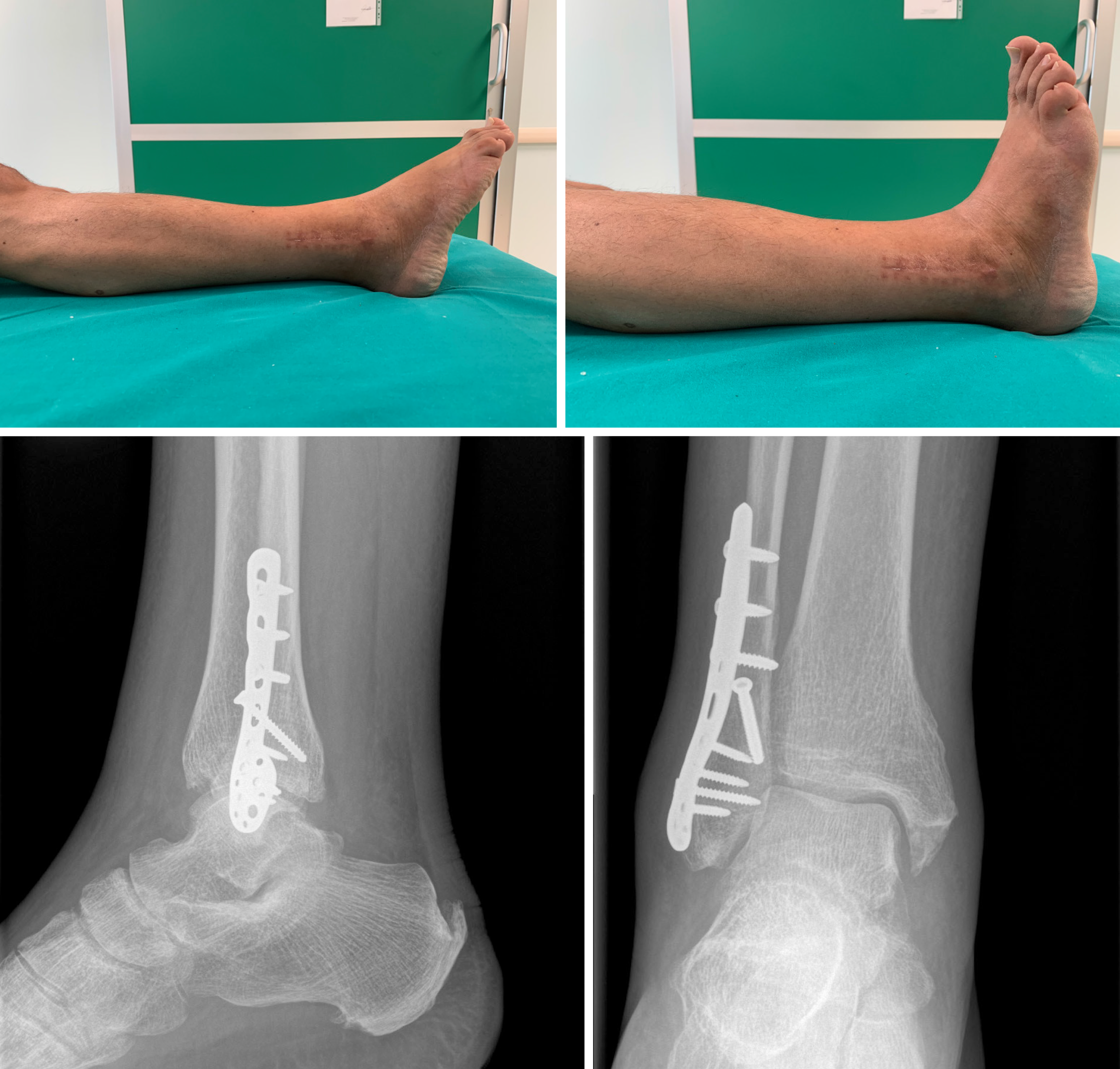
Figure 3 Radiographic and clinical results at 3 mo from a one-third tubular plate fixation of a Danis-Weber type B left distal fibula fracture.
Figure 4 Clinical and radiographic results at 3 mo from surgical treatment of a Danis-Weber type B right distal fibula fracture with an anatomic angular stable plate.
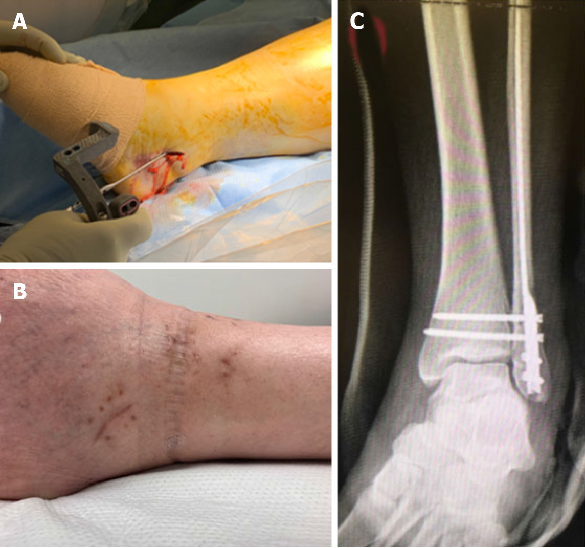
Figure 5 Clinical case of a Danis-Weber Type B left distal fibula fracture treated with locked nailing.
A: Intraoperative image demonstrating fibula nail insertion with guided instrumentation; B: Detail of the clinical result of minimally invasive approach for fibular nail insertion; C: Antero-posterior X-rays demonstrating fracture fixation with fibular nail completed with two guided intersyndesmotic screws.
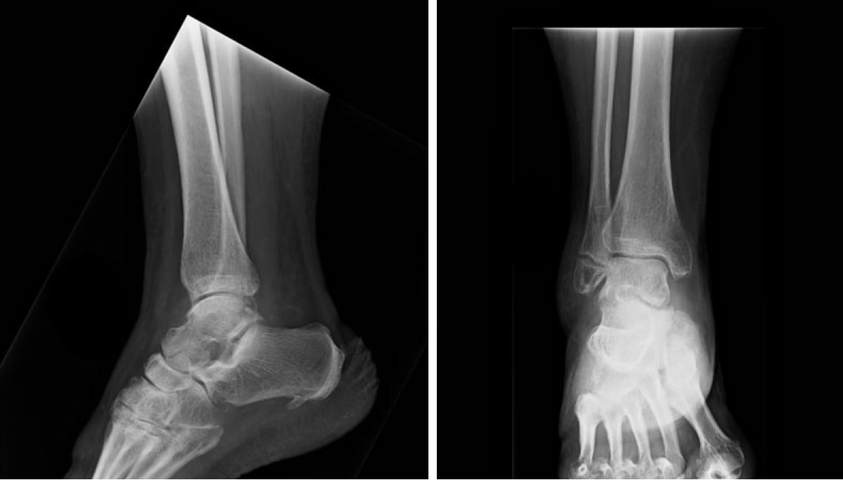
Figure 6 Lateral and antero-posterior view X-rays taken 4 mo after conservative treatment of a Danis-Weber type A distal fibula fracture.
Despite the radiographic evidence of nonunion the patient is completely asymptomatic, and no further treatment is indicated.
Figure 7 Clinical case of a Danis-Weber type C left distal fibula fracture treated with a one-third tubular plate.
At post-op X-rays, malreduction with residual medial displacement is demonstrated. A dedicated adjustable tibio-fibular suture button fixation was added to obtain anatomic reduction and correction of associated ankle instability.
- Citation: Canton G, Sborgia A, Maritan G, Fattori R, Roman F, Tomic M, Morandi MM, Murena L. Fibula fractures management. World J Orthop 2021; 12(5): 254-269
- URL: https://www.wjgnet.com/2218-5836/full/v12/i5/254.htm
- DOI: https://dx.doi.org/10.5312/wjo.v12.i5.254


