Copyright
©2014 Baishideng Publishing Group Inc.
World J Radiol. Nov 28, 2014; 6(11): 855-864
Published online Nov 28, 2014. doi: 10.4329/wjr.v6.i11.855
Published online Nov 28, 2014. doi: 10.4329/wjr.v6.i11.855
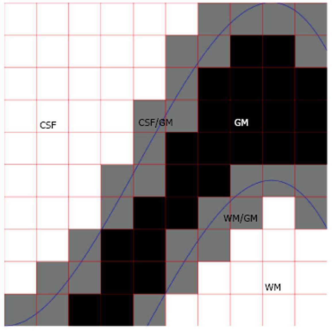
Figure 1 A schematic explanation of the partial volume effect in the context of brain magnetic resonance imaging.
Voxels composed of purely gray matter (GM) are colored in black color while voxels composed of cerebro-spinal fluid (CSF) or white matter (WM) are in white color. These are termed pure tissue voxels or pure voxels. Voxels composed of multiple tissue types, termed mixed voxels, are colored in gray. In the figure, these can be either voxels containing both CSF and GM tissue types or voxels containing both WM and GM tissue types. The actual anatomical boundaries between tissue types are shown in blue and red color is used to indicate voxel boundaries.
- Citation: Tohka J. Partial volume effect modeling for segmentation and tissue classification of brain magnetic resonance images: A review. World J Radiol 2014; 6(11): 855-864
- URL: https://www.wjgnet.com/1949-8470/full/v6/i11/855.htm
- DOI: https://dx.doi.org/10.4329/wjr.v6.i11.855


