Copyright
©The Author(s) 2016.
World J Radiol. Feb 28, 2016; 8(2): 200-209
Published online Feb 28, 2016. doi: 10.4329/wjr.v8.i2.200
Published online Feb 28, 2016. doi: 10.4329/wjr.v8.i2.200
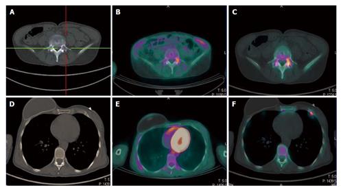
Figure 1 A 49-year-old breast cancer patient with bone relapse.
A lytic lesion on the fifth lumbar vertebra was detected with computed tomography in absence of local pain (A). The lesion showed high uptake both at 18F-FDG PET/CT (B) and 18F-NaF PET/CT (C), thus confirming its malignant nature. No further 18F-FDG avid metastasis was highlighted (E). By contrast a focal area of high 18F-NaF uptake was evident in the anterior branch of the seventh rib on the left side (F). This area was indeed corresponding to a small sclerotic indeterminate lesion on the CT and was considered as a further site of disease (D). The patient started systemic therapy rather than a targeted radiotherapy on the fifth lumbar vertebra. 18F-FDG: 2-deoxy-2-(18F)fluoro-D-glucose; 18F-NaF: 18F-sodium; PET/CT: Positron emission tomography/computed tomography.
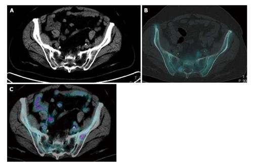
Figure 2 Evidence of the complementary features of 2-deoxy-2-(18F)fluoro-D-glucose and 18F-sodium positron emission tomography/computed tomography in the pelvis of the same breast cancer patient.
An 18F-NaF avid sclerotic lesion was detected in the right sacrum in absence of significant 18F-FDG uptake. By contrast high uptake of 18F-FDG was present in a small lytic lesion in the left iliac bone. Due to the absence of a significant local bone reaction, this small lesion did not show any uptake of 18F-NaF. Both lesions corresponded to metastatic sites of disease and disappeared after chemotherapy. 18F-FDG: 2-deoxy-2-(18F)fluoro-D-glucose; 18F-NaF: 18F-sodium; PET/CT: Positron emission tomography/computed tomography.
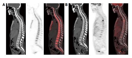
Figure 3 This area is likely to correspond to a bone marrow-confined metastasis not yet characterized by bone remodeling and thus falsely negative in both multidetector computed tomography and 18F-sodium positron emission tomography/computed tomography.
No structural lesions or area of 18F-NaF uptake are evident in the vertebral column of this breast cancer patient (A); by contrast an area of high focal 18F-FDG uptake was present in the sixth dorsal vertebra (B). 18F-NaF: 18F-sodium; 18F-FDG: 2-deoxy-2-(18F)fluoro-D-glucose.
- Citation: Capitanio S, Bongioanni F, Piccardo A, Campus C, Gonella R, Tixi L, Naseri M, Pennone M, Altrinetti V, Buschiazzo A, Bossert I, Fiz F, Bruno A, DeCensi A, Sambuceti G, Morbelli S. Comparisons between glucose analogue 2-deoxy-2-(18F)fluoro-D-glucose and 18F-sodium fluoride positron emission tomography/computed tomography in breast cancer patients with bone lesions. World J Radiol 2016; 8(2): 200-209
- URL: https://www.wjgnet.com/1949-8470/full/v8/i2/200.htm
- DOI: https://dx.doi.org/10.4329/wjr.v8.i2.200


