Copyright
©The Author(s) 2016.
World J Radiol. Feb 28, 2016; 8(2): 200-209
Published online Feb 28, 2016. doi: 10.4329/wjr.v8.i2.200
Published online Feb 28, 2016. doi: 10.4329/wjr.v8.i2.200
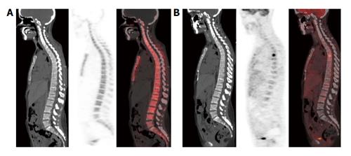
Figure 3 This area is likely to correspond to a bone marrow-confined metastasis not yet characterized by bone remodeling and thus falsely negative in both multidetector computed tomography and 18F-sodium positron emission tomography/computed tomography.
No structural lesions or area of 18F-NaF uptake are evident in the vertebral column of this breast cancer patient (A); by contrast an area of high focal 18F-FDG uptake was present in the sixth dorsal vertebra (B). 18F-NaF: 18F-sodium; 18F-FDG: 2-deoxy-2-(18F)fluoro-D-glucose.
- Citation: Capitanio S, Bongioanni F, Piccardo A, Campus C, Gonella R, Tixi L, Naseri M, Pennone M, Altrinetti V, Buschiazzo A, Bossert I, Fiz F, Bruno A, DeCensi A, Sambuceti G, Morbelli S. Comparisons between glucose analogue 2-deoxy-2-(18F)fluoro-D-glucose and 18F-sodium fluoride positron emission tomography/computed tomography in breast cancer patients with bone lesions. World J Radiol 2016; 8(2): 200-209
- URL: https://www.wjgnet.com/1949-8470/full/v8/i2/200.htm
- DOI: https://dx.doi.org/10.4329/wjr.v8.i2.200


