Copyright
©2014 Baishideng Publishing Group Inc.
World J Radiol. Nov 28, 2014; 6(11): 850-854
Published online Nov 28, 2014. doi: 10.4329/wjr.v6.i11.850
Published online Nov 28, 2014. doi: 10.4329/wjr.v6.i11.850
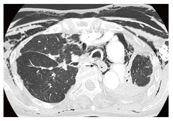
Figure 1 Chest computed tomography scan of an 82-year-old woman shows an injury to the posterior wall of the trachea, massive pneumomediastinum, and subcutaneous emphysema due to ruptured pars membranosa (arrow).
Figure 2 A 21-year-old woman with hypothyroidism and symptoms of cervical discomfort and tenderness.
Multidetector-row computed tomography scan demonstrates air collection along the perivascular connective tissue, the Macklin effect (arrows), in the peripheral area (A) and in the perihilar area (B), and pneumomediastinum. Reprinted from ref. [5].
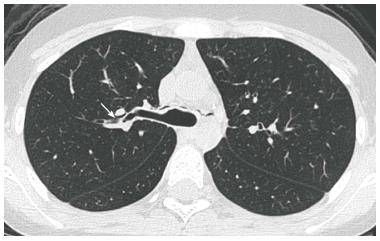
Figure 3 A 15-year-old girl with acute myeloid leukemia.
Multidetector-row computed tomography scan demonstrates air collection along the perivascular connective tissue and the Macklin effect (arrow) in the perihilar area. A small pneumomediastinum is also noted.
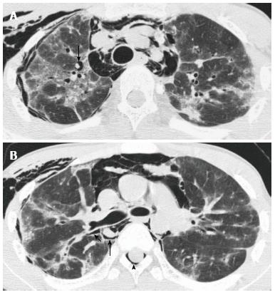
Figure 4 A 15-year-old girl with cryptogenic organizing pneumonia associated with graft-vs-host disease.
Multidetector-row computed tomography scan demonstrates air collection along the perivascular connective tissue, the Macklin effect (arrows) in the peripheral area (A) and in the perihilar area (B), and massive pneumomediastinum. This patient also has spinal pneumorrhachis (arrowhead). Reprinted from ref. [5].
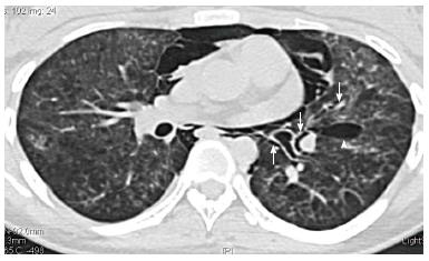
Figure 5 A 16-year-old girl with persistent spontaneous pneumomediastinum and pneumatocele.
Computed tomography shows massive pneumomediastinum and perihilar and peripheral Macklin effects (arrows). In the left lower lobe, a pneumatocele (arrowhead) is observed.
- Citation: Murayama S, Gibo S. Spontaneous pneumomediastinum and Macklin effect: Overview and appearance on computed tomography. World J Radiol 2014; 6(11): 850-854
- URL: https://www.wjgnet.com/1949-8470/full/v6/i11/850.htm
- DOI: https://dx.doi.org/10.4329/wjr.v6.i11.850


