Copyright
©2013 Baishideng.
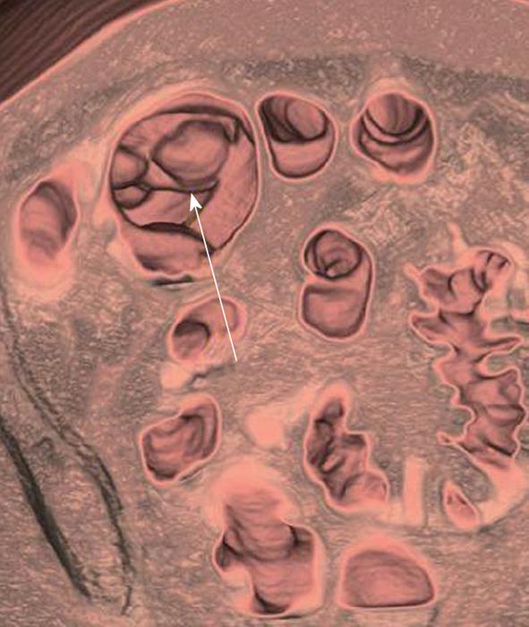
Figure 1 A 58-year-old man underwent computed tomography colonoscopy for a failed optical colonoscopy due to sigmoid stricture.
Computed tomography colonography was performed which showed a 1.5 cm cecal mass (arrow). Patient underwent right hemicolectomy and pathology confirmed cecal carcinoma. Failed optical colonoscopy is one of the most common indications for virtual colonoscopy.
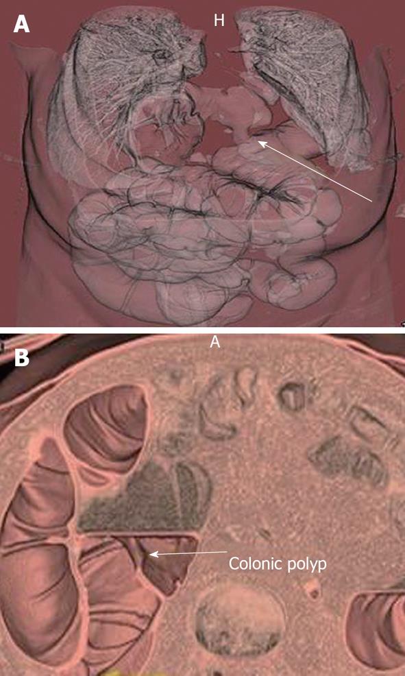
Figure 2 A 62-year-old man had a failed optical colonoscopy and underwent computed tomography colonoscopy.
A: 3D volume rendered image of computed tomography (CT) colonoscopy demonstrated a diaphragmatic hernia containing transverse colon (arrow), which was the reason for the failed optical colonoscopy; B: A 3 dimensional volume rendered image of the CT colonoscopy (B) also shows a 1.5 cm polyp (arrow) in the ascending colon.
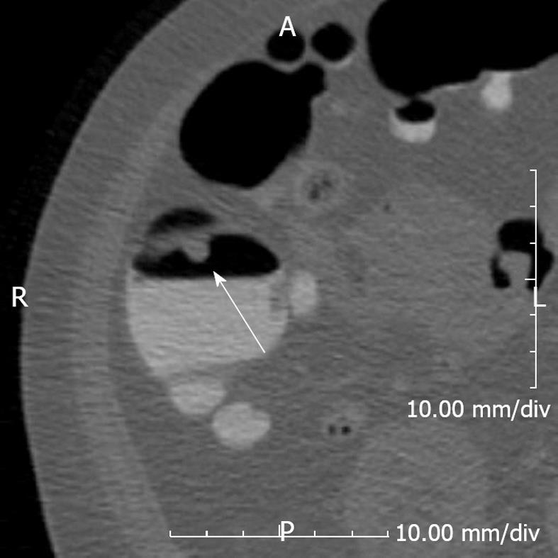
Figure 3 A 53-year-old female underwent computed tomography colonoscopy.
An 8 mm polyp (arrow) is seen in the ascending colon. Note the fluid in dependent position which potentially obscures pathology. Using different positions including supine, prone and decubitus positions causes fluid to shift position, thereby allowing a more thorough evaluation.
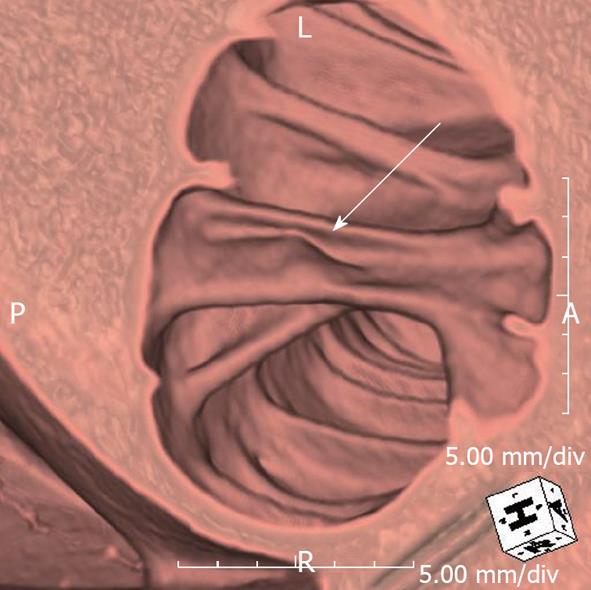
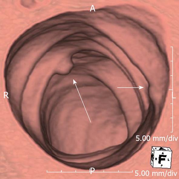
Figure 4 A 64-year-old man with nonspecific abdominal pain.
Computed tomography colonoscopy showed a 1 cm polyp (arrow) on the fold in ascending colon which was missed on the initial 2D review.
Figure 5 A 72-year-old male underwent computed tomography colonoscopy.
There is an 8 mm polyp in descending colon (long arrow), which is easily detected in the 3D view. Note that there is another ‘’polyp’’ like lesion (short arrow). This was shown to be a fecal material by correlating with the 2D view. False positives for polyp are one of the pitfalls of 3D review but this can be easily overcome by complementing 3D with 2D review.
- Citation: Ganeshan D, Elsayes KM, Vining D. Virtual colonoscopy: Utility, impact and overview. World J Radiol 2013; 5(3): 61-67
- URL: https://www.wjgnet.com/1949-8470/full/v5/i3/61.htm
- DOI: https://dx.doi.org/10.4329/wjr.v5.i3.61


