
















©2013 Baishideng.
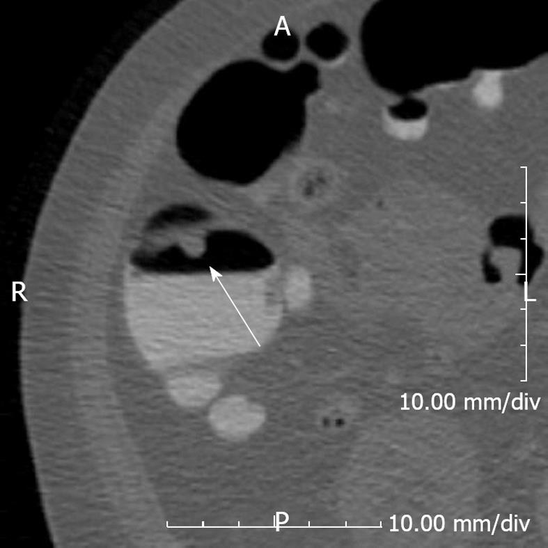
Figure 3 A 53-year-old female underwent computed tomography colonoscopy.
An 8 mm polyp (arrow) is seen in the ascending colon. Note the fluid in dependent position which potentially obscures pathology. Using different positions including supine, prone and decubitus positions causes fluid to shift position, thereby allowing a more thorough evaluation.
- Citation: Ganeshan D, Elsayes KM, Vining D. Virtual colonoscopy: Utility, impact and overview. World J Radiol 2013; 5(3): 61-67
- URL: https://www.wjgnet.com/1949-8470/full/v5/i3/61.htm
- DOI: https://dx.doi.org/10.4329/wjr.v5.i3.61