Copyright
©2013 Baishideng.
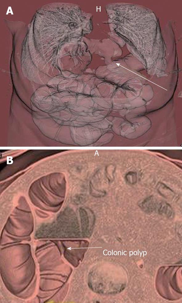
Figure 2 A 62-year-old man had a failed optical colonoscopy and underwent computed tomography colonoscopy.
A: 3D volume rendered image of computed tomography (CT) colonoscopy demonstrated a diaphragmatic hernia containing transverse colon (arrow), which was the reason for the failed optical colonoscopy; B: A 3 dimensional volume rendered image of the CT colonoscopy (B) also shows a 1.5 cm polyp (arrow) in the ascending colon.
- Citation: Ganeshan D, Elsayes KM, Vining D. Virtual colonoscopy: Utility, impact and overview. World J Radiol 2013; 5(3): 61-67
- URL: https://www.wjgnet.com/1949-8470/full/v5/i3/61.htm
- DOI: https://dx.doi.org/10.4329/wjr.v5.i3.61


