Copyright
©The Author(s) 2025.
World J Radiol. Jun 28, 2025; 17(6): 105632
Published online Jun 28, 2025. doi: 10.4329/wjr.v17.i6.105632
Published online Jun 28, 2025. doi: 10.4329/wjr.v17.i6.105632
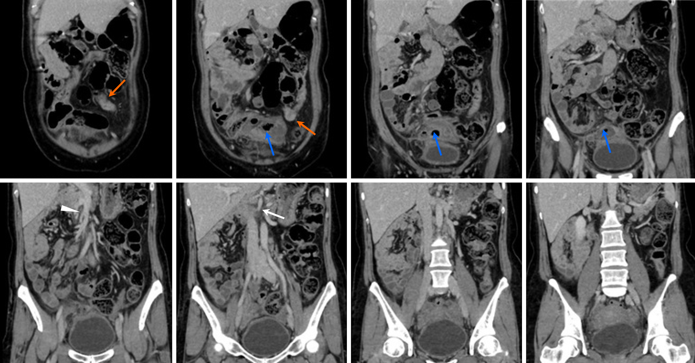
Figure 1 Contrast-enhanced coronal computed tomography images of a 34-year-old woman at 19 weeks gestation presenting with right lower quadrant abdominal pain.
Images demonstrate an enlarged appendix (approximately 7 mm in diameter) with fecal stones located adjacent to the gravid uterus (orange arrow), consistent with acute appendicitis in pregnancy (blue arrow).
Figure 2 Contrast-enhanced coronal computed tomography images of a 57-year-old male with left lower quadrant abdominal pain demonstrating situs inversus totalis.
The liver and appendix are visualized in the left abdomen. The appendix is swollen at its root (approximately 1 cm in diameter) with high-density fecal material, indicative of acute appendicitis associated with complete visceral inversion. Orange arrow: Inflamed appendix arising from the cecum in the left lower quadrant. The appendix is filled with contrast and shows distal swelling up to 9 mm in diameter; Blue arrow: Inverted position of the liver on the left side of the abdomen, indicating complete visceral inversion.
Figure 3 Contrast-enhanced coronal computed tomography images of a 44-year-old woman presenting with lower abdominal pain and fever, showing evidence of intestinal malrotation with abnormal positioning of the superior mesenteric artery (white triangle) and supe
- Citation: Asahi K. Retrospective analysis of computed tomography examinations in patients with lower abdominal pain: A single-center experience. World J Radiol 2025; 17(6): 105632
- URL: https://www.wjgnet.com/1949-8470/full/v17/i6/105632.htm
- DOI: https://dx.doi.org/10.4329/wjr.v17.i6.105632


