
















Copyright: ©Author(s) 2026.
World J Gastrointest Surg. Nov 27, 2026; 18(11): 125637
Published online Nov 27, 2026. doi: 10.4240/wjgs.125637
Published online Nov 27, 2026. doi: 10.4240/wjgs.125637
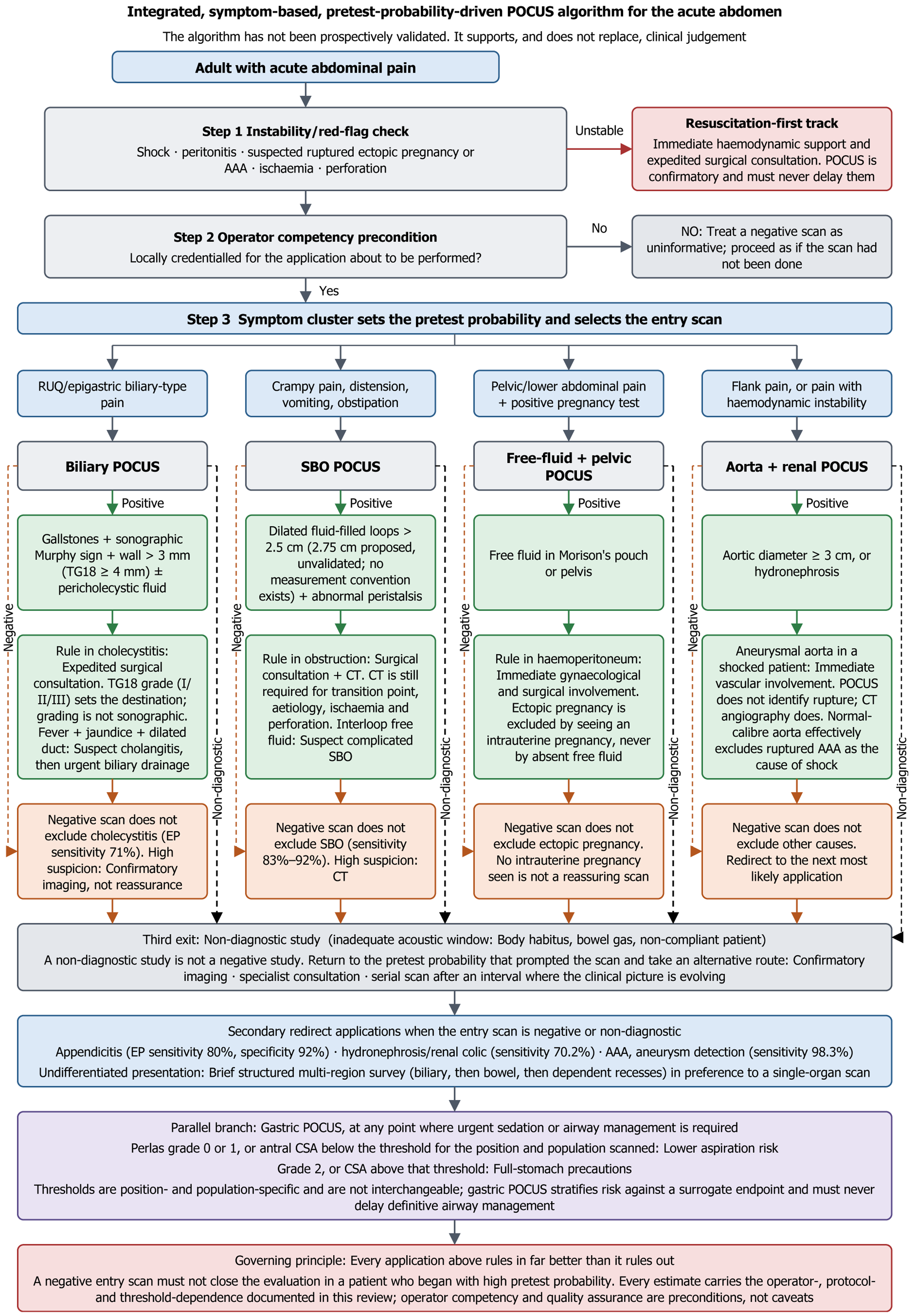
Figure 1 Integrated, symptom-based, pretest-probability-driven point-of-care ultrasound scanning algorithm for the acute abdomen.
The pathway opens with two entry conditions: A mandatory instability and red-flag check (red) – shock, peritonitis, suspected ruptured ectopic pregnancy, ruptured abdominal aortic aneurysm, bowel ischaemia or perforation divert the patient to a resuscitation-first track with expedited surgical consultation, and bedside scanning never delays it – and an operator-competency precondition, below which a negative scan is treated as uninformative. For the stable patient, each presenting symptom cluster sets the pretest probability and selects an entry scan. Every scan node has three exits: Adequate and positive (green, rule-in actions), adequate and negative (orange, rule-out caution and redirect), and non-diagnostic (grey), which returns the clinician to the pretest probability and mandates confirmatory imaging, consultation or a serial scan. A rule-in biliary scan routes to surgical consultation with Tokyo Guidelines 2018 severity grading, which determines the destination, and the cholangitis pathway is shown separately. The aortic branch distinguishes detection of an aneurysmal aorta from detection of rupture. A parallel gastric branch (purple) stratifies aspiration risk at any point. The algorithm has not been prospectively validated. POCUS: Point-of-care ultrasound; AAA: Abdominal aortic aneurysm; RUQ: Right upper quadrant; TG18: Tokyo Guidelines 2018; CT: Computed tomography; CSA: Cross-sectional area.
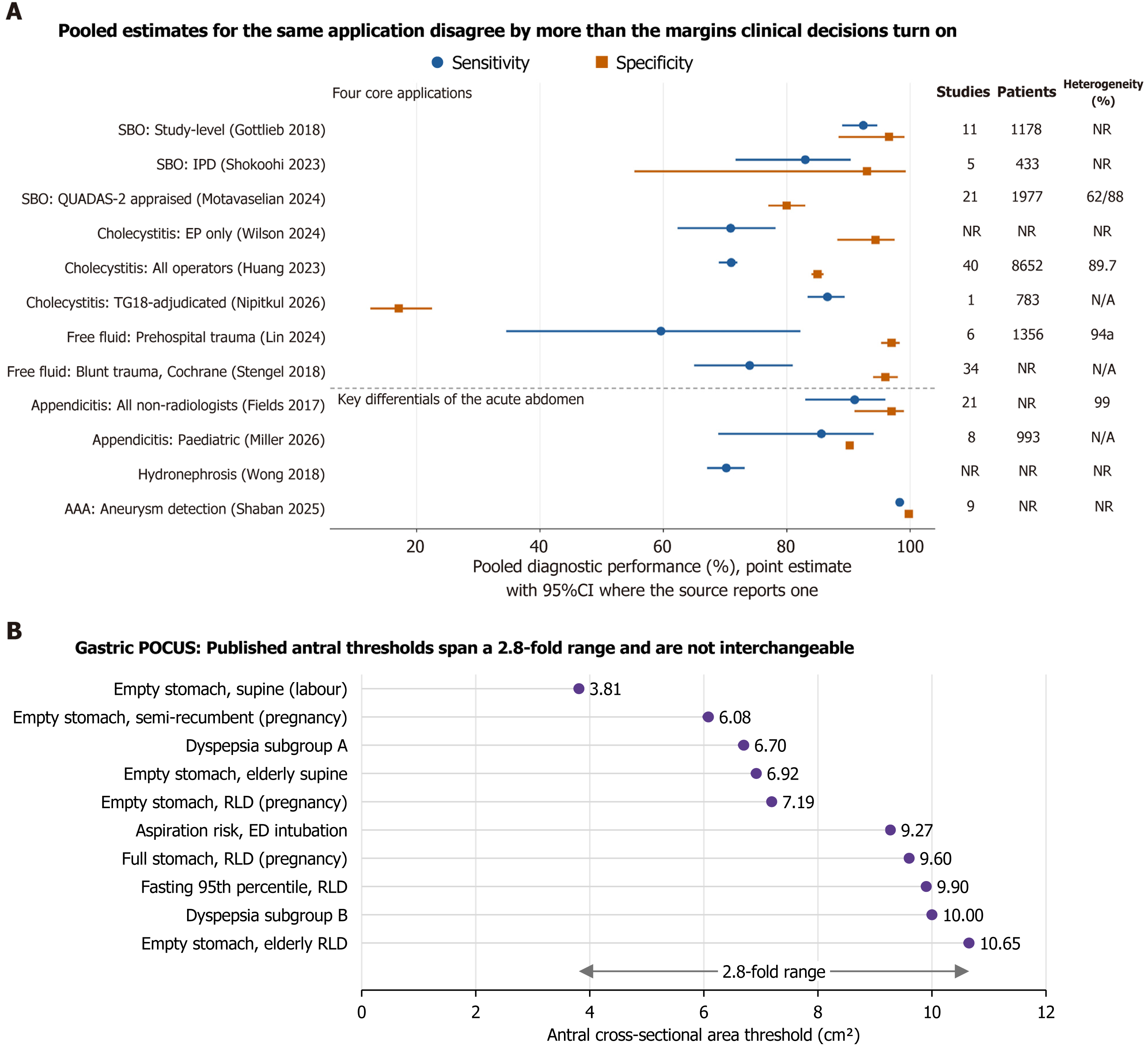
Figure 2 Pooled diagnostic accuracy, heterogeneity and threshold dispersion in emergency point-of-care ultrasound.
A: Circles denote pooled sensitivity and squares pooled specificity, with 95%CIs where the source reports them. The four core applications appear above the divider and the key differentials of the acute abdomen below it. Where more than one meta-analysis exists for an application, each source is plotted separately rather than combined, so that between-source dispersion is visible. The right-hand columns give the number of contributing studies and patients and the heterogeneity statistic reported by each source; values of I² at or above 60% are shown in orange, and NR indicates that the value could not be recovered from the retrievable source text; B: Published antral cross-sectional area thresholds for gastric point-of-care ultrasound plotted on a common scale. Gastric assessment is shown separately because it stratifies aspiration risk against a surrogate endpoint rather than functioning as a diagnostic accuracy test. Each threshold is specific to the body position, population and target volume in which it was derived, and the thresholds are not interchangeable. SBO: Small-bowel obstruction; IPD: Individual-patient-data; QUADAS-2: Quality Assessment of Diagnostic Accuracy Studies 2; EP: Emergency physician; TG18: Tokyo Guidelines 2018; AAA: Abdominal aortic aneurysm; RLD: Right lateral decubitus; POCUS: Point-of-care ultrasound; N/A: Not applicable.
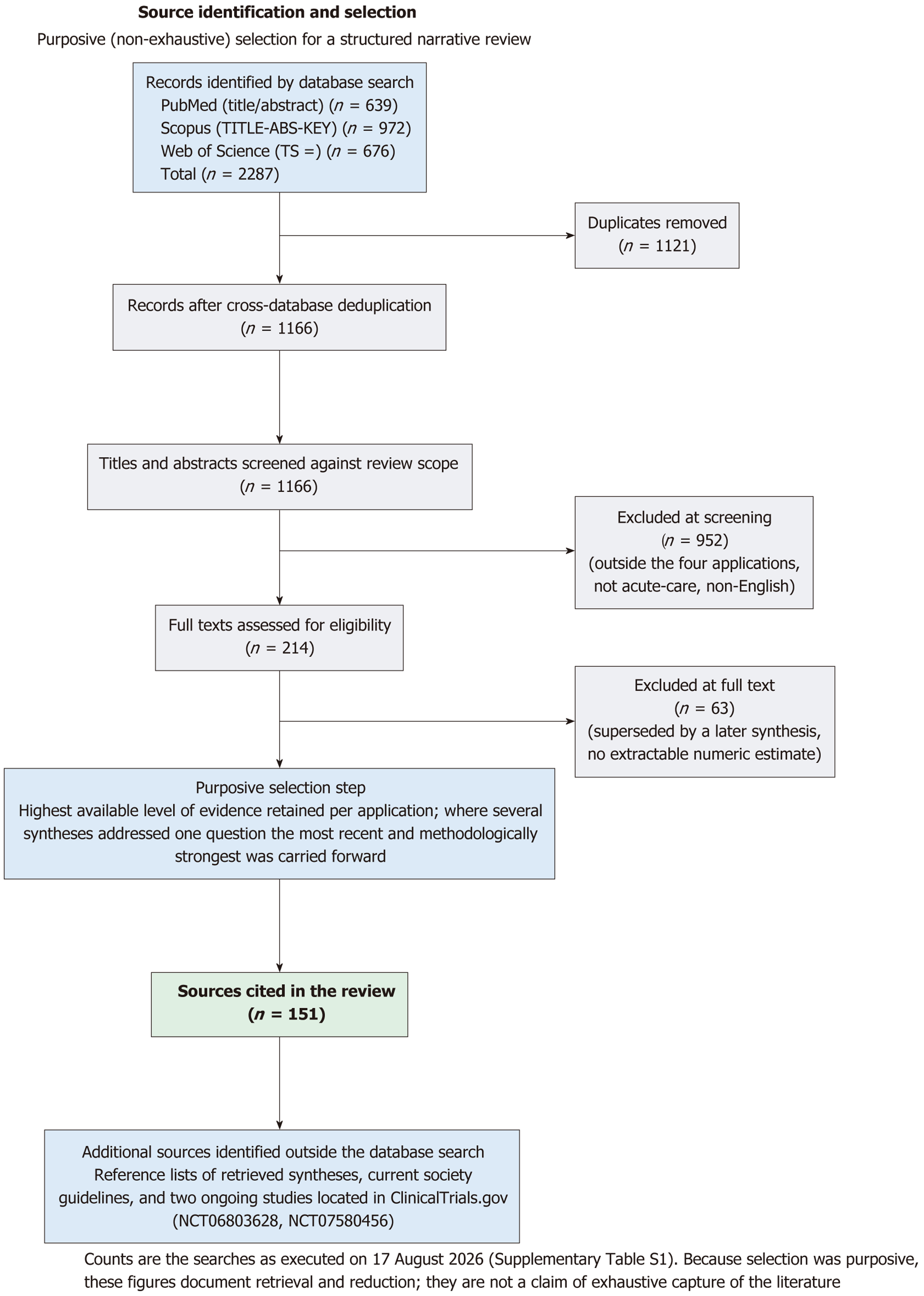
Figure 3 Source identification and selection for this structured narrative review.
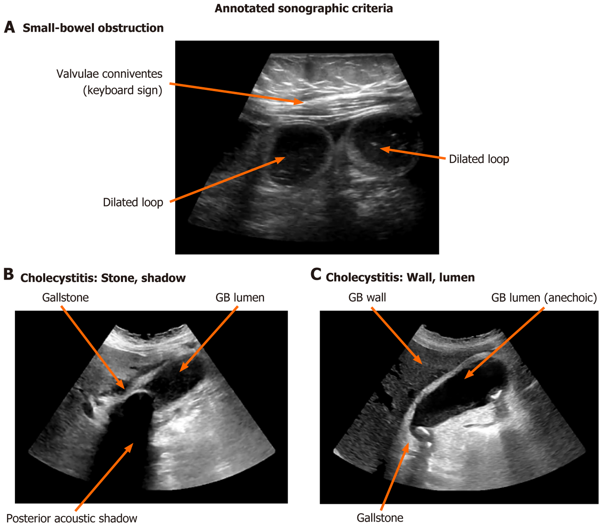
Figure 4 Annotated sonographic criteria on representative point-of-care ultrasound images acquired by the authors.
A: Small-bowel obstruction – a fluid-filled dilated loop with valvulae conniventes (the keyboard sign), with a second dilated loop also labelled; B: Acute cholecystitis – an intraluminal gallstone with posterior acoustic shadowing, adjacent to the anechoic gallbladder lumen; C: Acute cholecystitis – the gallbladder wall, the anechoic lumen and a gallstone. Arrows indicate the labelled structures. Diagnostic thresholds (dilated small-bowel loop > 2.5 cm conventionally, with 2.75 cm proposed as a bedside-specific candidate; gallbladder wall > 3 mm in the emergency literature and ≥ 4 mm in the Tokyo Guidelines 2018) are defined, referenced and compared in the main text. GB: Gallbladder.
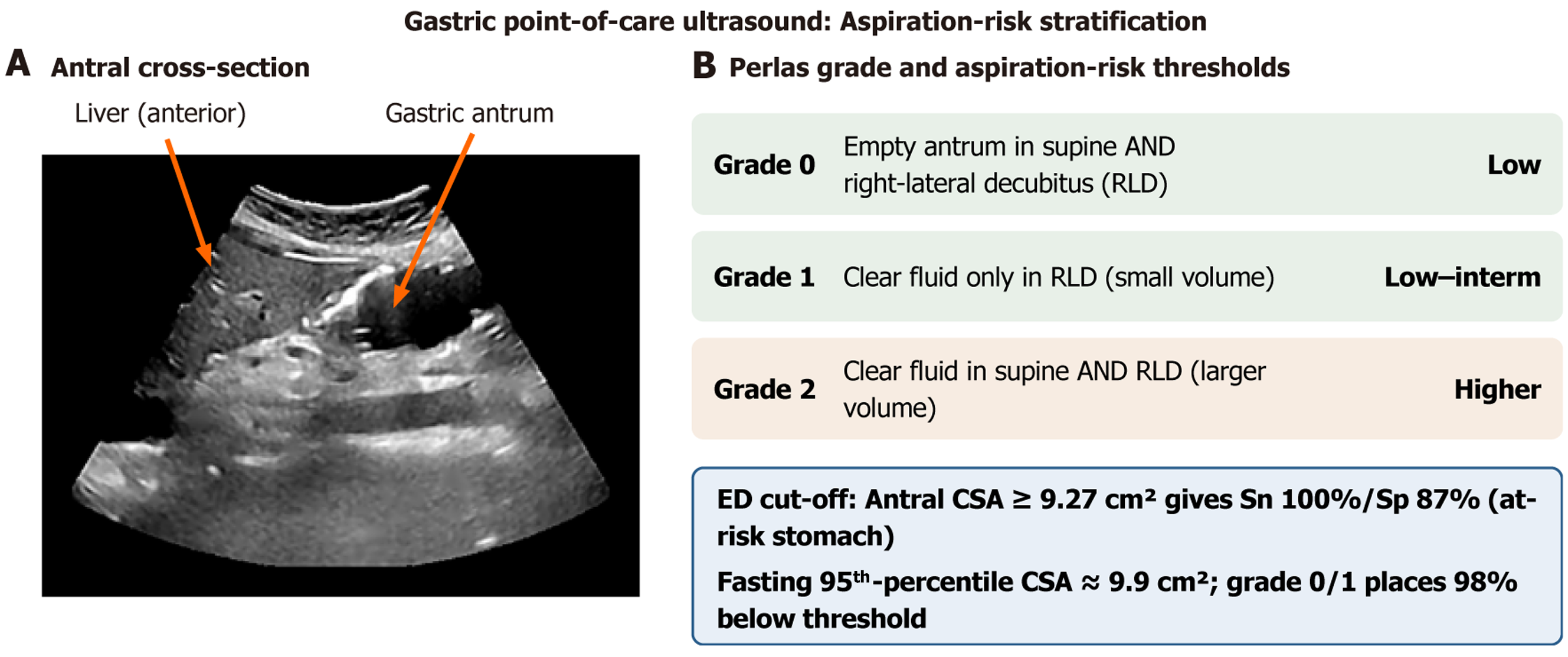
Figure 5 Gastric point-of-care ultrasound for aspiration-risk stratification.
A: Representative antral image acquired by the authors, showing the gastric antrum with the anterior liver as the acoustic window; antral cross-sectional area is obtained from the anteroposterior and craniocaudal diameters as described in the text; B: The Perlas qualitative grading system (grades 0-2) with the corresponding aspiration-risk category, and the emergency-department and fasting-reference antral cross-sectional area cut-offs. Each threshold applies only to the body position and population in which it was derived, and the full set of published thresholds and their dispersion is shown in Figure 2B. All cited studies relate antral measurement to gastric volume; none relates it to aspiration or to any other patient-centred outcome. RLD: Right lateral decubitus.
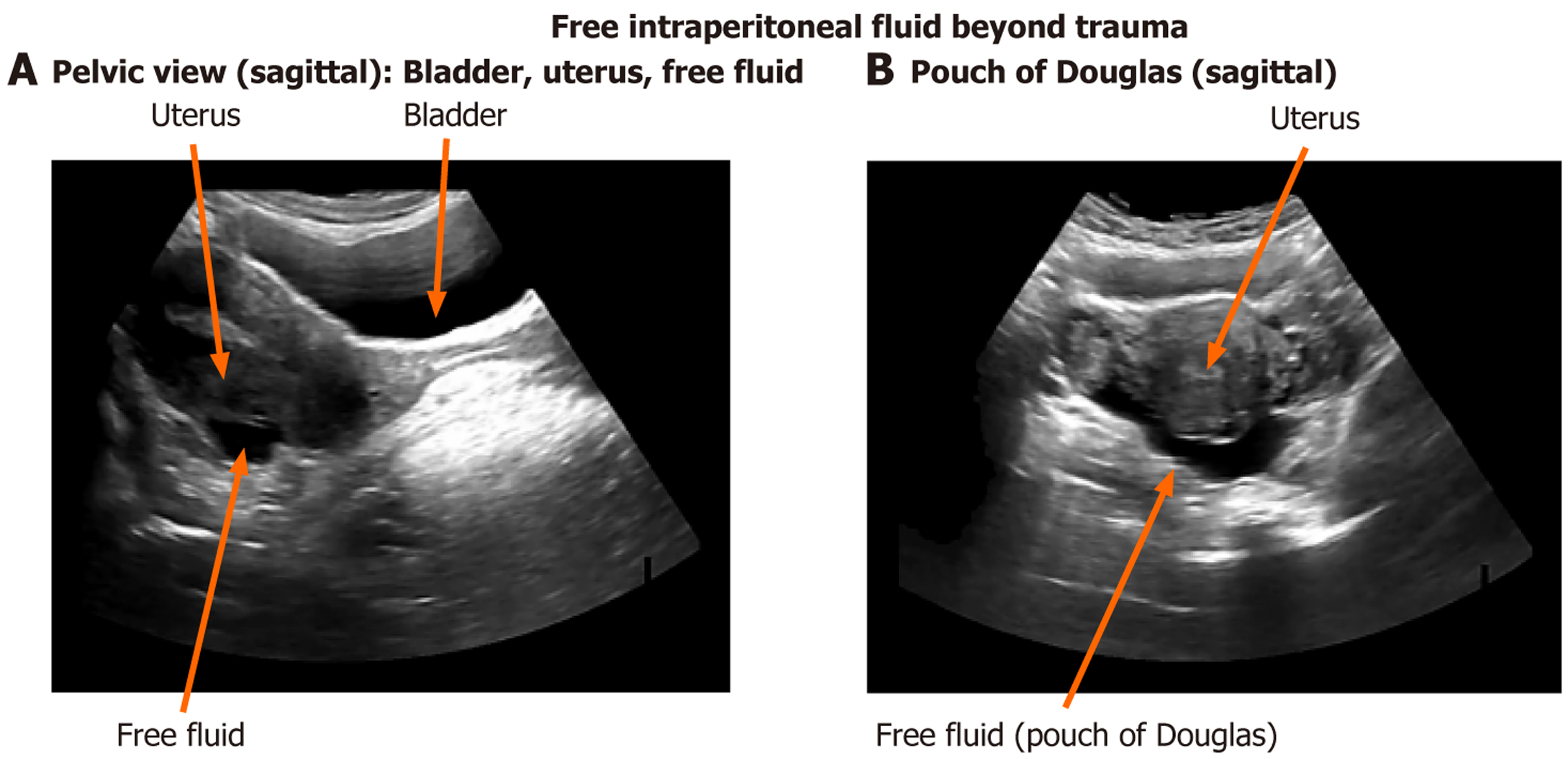
Figure 6 Point-of-care ultrasound of free intraperitoneal fluid beyond trauma, on images acquired by the authors.
A: Sagittal pelvic view showing the urinary bladder, the uterus and anechoic free fluid in the dependent pelvis; B: Sagittal pelvic view showing free fluid in the rectouterine pouch (pouch of Douglas) posterior to the uterus. Arrows indicate the labelled structures. In a high-pretest-probability obstetric population, even a small anechoic collection in a dependent recess is clinically meaningful; the significance of free fluid in other populations is interpreted in its own clinical context.
- Citation: Danış F, Kudu E. Point-of-care ultrasound in the acute abdomen and gastrointestinal emergencies: An evidence-based guide and the case for standardisation. World J Gastrointest Surg 2026; 18(11): 125637
- URL: https://www.wjgnet.com/1948-9366/full/v18/i11/125637.htm
- DOI: https://dx.doi.org/10.4240/wjgs.125637