
















Copyright: ©Author(s) 2026.
World J Gastrointest Surg. Nov 27, 2026; 18(11): 125637
Published online Nov 27, 2026. doi: 10.4240/wjgs.125637
Published online Nov 27, 2026. doi: 10.4240/wjgs.125637
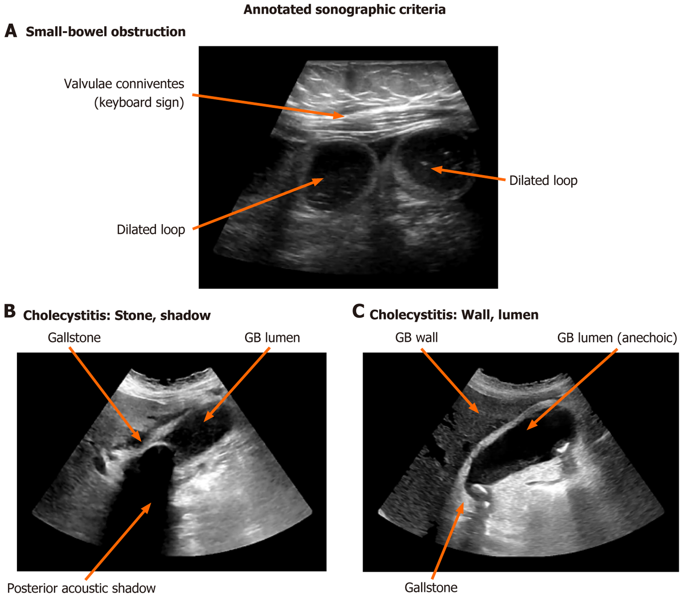
Figure 4 Annotated sonographic criteria on representative point-of-care ultrasound images acquired by the authors.
A: Small-bowel obstruction – a fluid-filled dilated loop with valvulae conniventes (the keyboard sign), with a second dilated loop also labelled; B: Acute cholecystitis – an intraluminal gallstone with posterior acoustic shadowing, adjacent to the anechoic gallbladder lumen; C: Acute cholecystitis – the gallbladder wall, the anechoic lumen and a gallstone. Arrows indicate the labelled structures. Diagnostic thresholds (dilated small-bowel loop > 2.5 cm conventionally, with 2.75 cm proposed as a bedside-specific candidate; gallbladder wall > 3 mm in the emergency literature and ≥ 4 mm in the Tokyo Guidelines 2018) are defined, referenced and compared in the main text. GB: Gallbladder.
- Citation: Danış F, Kudu E. Point-of-care ultrasound in the acute abdomen and gastrointestinal emergencies: An evidence-based guide and the case for standardisation. World J Gastrointest Surg 2026; 18(11): 125637
- URL: https://www.wjgnet.com/1948-9366/full/v18/i11/125637.htm
- DOI: https://dx.doi.org/10.4240/wjgs.125637