
















Copyright: ©Author(s) 2026.
World J Gastrointest Surg. Nov 27, 2026; 18(11): 125637
Published online Nov 27, 2026. doi: 10.4240/wjgs.125637
Published online Nov 27, 2026. doi: 10.4240/wjgs.125637
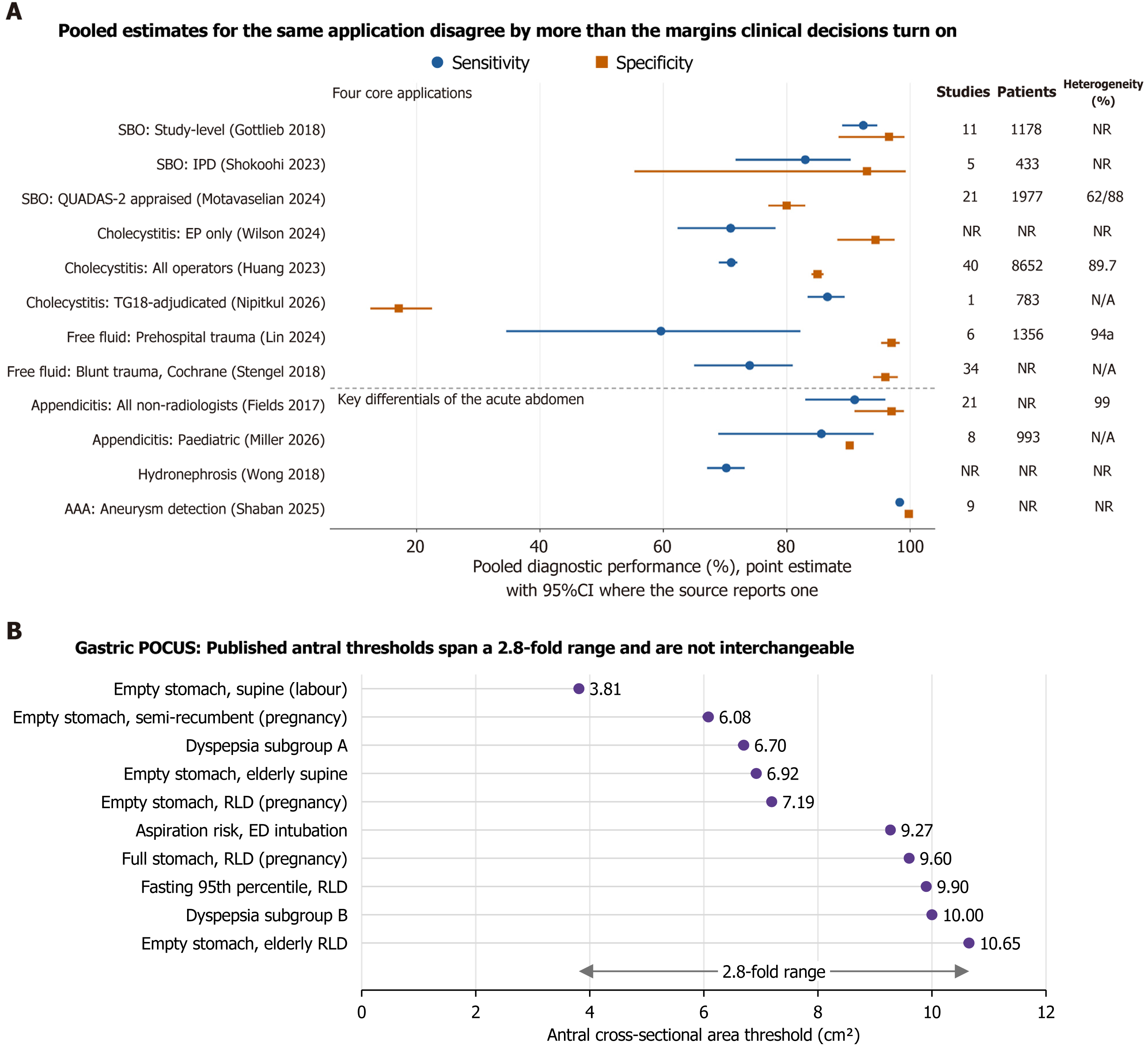
Figure 2 Pooled diagnostic accuracy, heterogeneity and threshold dispersion in emergency point-of-care ultrasound.
A: Circles denote pooled sensitivity and squares pooled specificity, with 95%CIs where the source reports them. The four core applications appear above the divider and the key differentials of the acute abdomen below it. Where more than one meta-analysis exists for an application, each source is plotted separately rather than combined, so that between-source dispersion is visible. The right-hand columns give the number of contributing studies and patients and the heterogeneity statistic reported by each source; values of I² at or above 60% are shown in orange, and NR indicates that the value could not be recovered from the retrievable source text; B: Published antral cross-sectional area thresholds for gastric point-of-care ultrasound plotted on a common scale. Gastric assessment is shown separately because it stratifies aspiration risk against a surrogate endpoint rather than functioning as a diagnostic accuracy test. Each threshold is specific to the body position, population and target volume in which it was derived, and the thresholds are not interchangeable. SBO: Small-bowel obstruction; IPD: Individual-patient-data; QUADAS-2: Quality Assessment of Diagnostic Accuracy Studies 2; EP: Emergency physician; TG18: Tokyo Guidelines 2018; AAA: Abdominal aortic aneurysm; RLD: Right lateral decubitus; POCUS: Point-of-care ultrasound; N/A: Not applicable.
- Citation: Danış F, Kudu E. Point-of-care ultrasound in the acute abdomen and gastrointestinal emergencies: An evidence-based guide and the case for standardisation. World J Gastrointest Surg 2026; 18(11): 125637
- URL: https://www.wjgnet.com/1948-9366/full/v18/i11/125637.htm
- DOI: https://dx.doi.org/10.4240/wjgs.125637